Courtesy: Prof Nabil Ebraheim, University of Toledo, USA
Introduction
The sternoclavicular joint (SCJ) is the only true bony articulation connecting the upper limb to the axial skeleton. Although small, it plays a critical role in shoulder girdle motion, enabling full upper-extremity function.
Anterior dislocation of the SCJ is rare but clinically important, primarily due to the joint’s close anatomical relationship with vital mediastinal structures. While anterior dislocations are generally less dangerous than posterior dislocations, they can still lead to pain, cosmetic deformity, instability, and functional limitation if not managed appropriately.
Anatomical Considerations
Joint Type and Structure
The sternoclavicular joint is a saddle-type synovial joint formed by:
-
Medial end of the clavicle
-
Manubrium of the sternum
-
First costal cartilage
Despite its saddle configuration, the SCJ functions biomechanically like a ball-and-socket joint, allowing a wide range of motion.
Ligamentous Stabilizers
The stability of the SCJ depends largely on strong ligamentous support rather than bony congruity:
-
Anterior and Posterior Sternoclavicular Ligaments
Reinforce the joint capsule and resist translation. -
Interclavicular Ligament
Connects the medial ends of both clavicles across the sternum and limits excessive depression. -
Costoclavicular Ligament (CC ligament)
Anchors the clavicle to the first rib; the primary restraint against excessive elevation and displacement. -
Intra-articular Disc
A fibrocartilaginous structure dividing the joint into two compartments, acting as a shock absorber and improving congruity.
Muscular Support
Dynamic stabilization is provided by surrounding muscles:
-
Subclavius – Provides direct support beneath the clavicle
-
Sternocleidomastoid and pectoralis major – Contribute secondary stability and influence movement patterns
Range of Motion
The SCJ permits multiplanar motion essential for shoulder mechanics:
-
Elevation / Depression: ~30–35°
-
Protraction / Retraction
-
Axial rotation: ~45°
These movements are critical for overhead activity, reaching, and cross-body motions.
Surrounding Structures (Clinical Relevance)
The SCJ lies in close proximity to vital structures, including:
-
Brachiocephalic vein
-
Subclavian vessels
-
Trachea
-
Esophagus
This anatomical relationship explains why posterior dislocations can be life-threatening, whereas anterior dislocations, though more common, are typically less dangerous but still clinically significant.
Mechanism of Injury
SCJ dislocations are broadly classified based on etiology into:
-
Traumatic anterior dislocation
-
Atraumatic (spontaneous) dislocation
1. Traumatic Anterior SCJ Dislocation
Mechanism:
-
High-energy blunt trauma, commonly seen in:
-
Contact sports (football, wrestling)
-
Motor vehicle accidents
-
Falls onto the shoulder
-
Biomechanics:
-
A posterolateral force to the shoulder drives the lateral clavicle posteriorly
-
This causes the medial clavicle to pivot anteriorly
-
Resultant tearing of stabilizing ligaments leads to dislocation
Structures Disrupted:
-
Sternoclavicular ligaments
-
Interclavicular ligament
-
Costoclavicular ligament
2. Atraumatic (Spontaneous) Dislocation
Epidemiology & Causes:
-
More common in young individuals
-
Associated with:
-
Ligamentous laxity
-
Repetitive overhead activity
-
Connective tissue disorders
-
Muscle imbalance
-
These dislocations often occur without a clear traumatic event and may be recurrent.
Clinical Presentation
Patients may present with:
-
Visible anterior prominence over the SCJ
-
Localized pain and tenderness
-
Instability or “popping” sensation
-
Reduced shoulder range of motion
-
Cosmetic concern
Neurological or vascular symptoms are rare in anterior dislocations but must always be assessed.
Imaging Evaluation
-
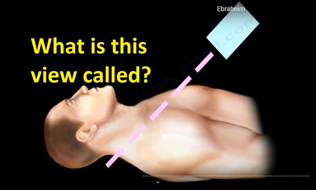
Plain radiographs (serendipity view) – Initial assessment
-
CT scan – Gold standard for confirming direction of dislocation and assessing surrounding structures
-
MRI – Useful in chronic or atraumatic cases to evaluate soft-tissue integrity
Management of Anterior SCJ Dislocation
Non-Operative Management (Mainstay of Treatment)
Most anterior SCJ dislocations are treated conservatively, even when reduction is incomplete.
Phase 1: Acute Phase (0–4 weeks)
Goals:
-
Pain control
-
Joint protection
-
Prevent further displacement
Management:
-
Sling immobilization
-
Analgesics and anti-inflammatory medication
-
Activity restriction:
-
No lifting, pushing, or pulling
-
Avoid shoulder elevation beyond 60°
-
-
Encourage scapular protraction (rounded shoulder posture) to reduce stress on the SCJ
Phase 2: Early Mobilization (4–8 weeks)
Goals:
-
Restore gentle motion
-
Prevent stiffness and adhesions
Rehabilitation:
-
Passive range-of-motion exercises:
-
Pendulum exercises
-
Wall crawls
-
-
Emphasis on:
-
Scapular mobility
-
Postural correction
-
Soft-tissue mobilization
-
?? Avoid excessive abduction, flexion, or loading of the joint.
Phase 3: Functional Rehabilitation (8–12 weeks)
Progression to:
-
Active-assisted ? active ROM
-
Isometric strengthening of:
-
Rotator cuff
-
Scapular stabilizers (serratus anterior, rhomboids, trapezius)
-
Monitoring for:
-
Pain
-
Clicking or prominence
-
Recurrent instability
Phase 4: Return to Activity (3–6 months)
Gradual return to:
-
Overhead activities
-
Resistance training
-
Sport-specific movements
Return to contact sports is typically allowed after 4–6 months, provided:
-
Full, pain-free ROM
-
Symmetric strength
-
Imaging confirms joint stability and healing
Surgical Management (Rare)
Surgery is rarely required for anterior SCJ dislocation and is reserved for:
-
Persistent pain
-
Recurrent symptomatic instability
-
Significant functional limitation despite adequate conservative care
Key Take-Home Points
-
The SCJ is a small but functionally critical joint
-
Anterior dislocations are more common and usually benign
-
Conservative treatment yields excellent functional outcomes
-
Rehabilitation should focus on posture, scapular control, and gradual loading
-
Surgery is the exception, not the rule
Leave a Reply