
Courtesy: Prof Nabil Ebraheim, University of Toledo, Ohio, USA
Cervical Spine & Upper Extremity Examination
Overview
The cervical spine examination follows the usual orthopaedic sequence:
Inspection – Palpation – Range of motion – Strength – Neurovascular examination – Specific tests – Gait
The aim is not only to identify cervical pathology, but also to distinguish it from shoulder and peripheral nerve disorders.
- Inspection
Inspect the patient for:
- Cervical alignment and deformity in coronal and sagittal planes
- Shoulder imbalance
- Scapular winging
- Rib prominence
- Surgical scars
- Cervical spine
- Shoulder
- Carpal tunnel
- Cubital tunnel
- Skin abnormalities, including café-au-lait spots in neurofibromatosis
- Muscle wasting/atrophy
The speaker gives normal sagittal alignment as:
- Cervical lordosis: 20–40°
- Thoracic kyphosis: 20–45°
Muscle Wasting
Deltoid + biceps wasting may suggest C5–C6 pathology.
Also look for:
- Abducted little finger — Wartenberg sign
- First dorsal interosseous wasting
These may indicate ulnar nerve dysfunction/hand muscle weakness in the speaker’s discussion.
- Palpation
Palpate systematically from proximal to distal.
Bony structures
Start from the:
Occiput – cervical spinous processes
Assess for:
- Asymmetry
- Local tenderness
Soft tissues
Palpate:
- Paraspinal muscles
- Scapula
- Ribs
- Trapezius
- Rhomboids
- Levator scapulae
- Trigger points/spasm
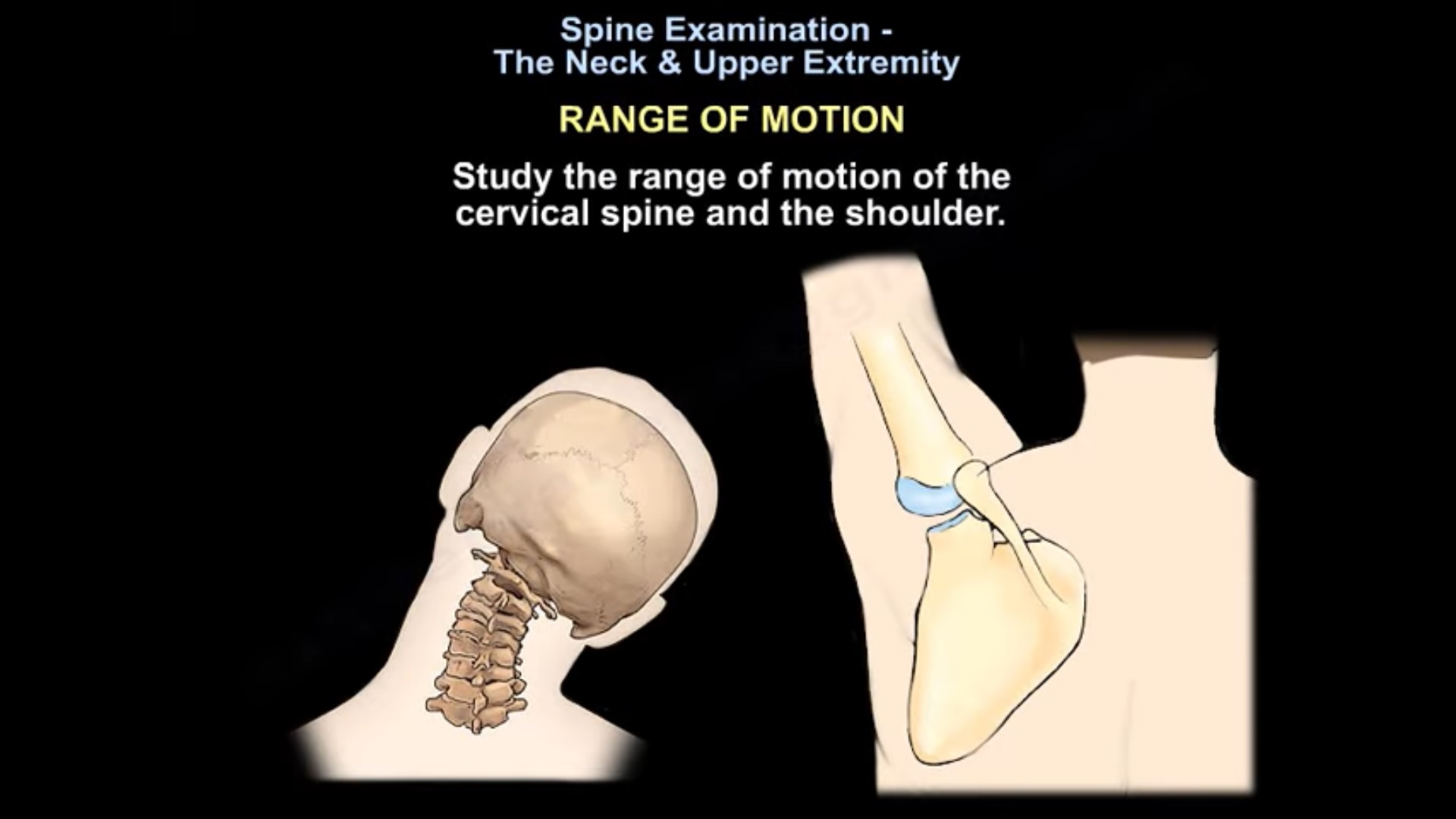
- Cervical Spine Range of Motion
Assess:
| Movement | ROM stated by speaker |
| Flexion | ~45° |
| Extension | ~70° |
| Lateral bending | ~45° |
| Rotation | ~75° |
During flexion, the speaker notes that the chin approaches/touches the chest.
A shoulder examination should also be performed.
- Provocative Tests
The transcript discusses:
- Spurling test
- Hoffmann sign
- Lhermitte sign
- Shoulder abduction test
- Shoulder impingement testing
- Carpal tunnel compression test
- Tinel sign at the cubital tunnel
These help differentiate cervical spine disease from shoulder and peripheral nerve pathology.
- Spurling Test
Purpose
Assess for cervical nerve-root irritation/compression.
Technique
- Patient seated.
- Turn the head toward the affected side.
- Examiner applies downward compression over the head.
Positive Test
Radiating pain down the patient’s arm produced by cervical compression.
This supports cervical radicular pathology.
Viva
Head toward symptomatic side + axial compression – radiating arm pain = positive Spurling test.
- Hoffmann Sign
Technique
The examiner flicks the nail of the:
- Middle finger, or
- Ring finger
A positive response produces flexion involving the index finger/thumb region, as described in the transcript.
Significance
Positive Hoffmann sign suggests an upper motor neuron lesion associated with spinal cord compression.
- Lhermitte Sign
Also called the Lhermitte phenomenon.
Positive Finding
Flexion/movement of the neck produces:
An electric shock-like sensation radiating down the spine.
It may extend into:
- Legs
- Arms
- Occasionally the trunk
Quick Recall
Neck flexion – electric shock down spine = Lhermitte sign.
- Shoulder Abduction Test
Technique
Ask the patient to:
Place the hand over the head, abducting the shoulder.
Positive Test
The patient’s radicular symptoms are relieved.
According to the speaker, relief occurs because shoulder abduction:
Decreases tension on the nerve roots.
Therefore, relief favors cervical nerve-root irritation rather than intrinsic shoulder pathology.
Exam Pearl
Shoulder abduction RELIEVES radicular pain – cervical nerve-root pathology.
- Carpal Tunnel Compression Test
The transcript refers to the carpal tunnel compression test.
Technique
- Apply even pressure using both thumbs directly over the median nerve at the carpal tunnel.
- Maintain for approximately 30 seconds.
Positive Test
Reproduction of symptoms in the:
Median nerve distribution
supports carpal tunnel syndrome.
The speaker describes this as a highly sensitive test.
- Tinel Sign at the Elbow
Tap over the ulnar nerve at the cubital tunnel.
Reproduction of ulnar nerve symptoms suggests:
Ulnar nerve entrapment at the cubital tunnel.
- Gait Examination
Gait should not be forgotten during cervical spine examination.
The transcript associates a:
Wide-based/shuffling gait with neurological disease and myelopathy.
It also mentions gait disturbance associated with foot drop.
- Neurological Examination
The neurological examination includes:
Motor + Sensory + Reflexes + Upper motor neuron signs
The speaker recommends testing at least one key muscle from each C5–T1 nerve-root group.
- Manual Muscle Power
The transcript uses the 0–5 grading system:
| Grade | Finding |
| 0 | No evidence of contraction |
| 1 | Minimal contraction |
| 2 | Movement with gravity eliminated |
| 3 | Movement against gravity |
| 4 | Movement against some resistance |
| 5 | Normal power |
- Cervical Root Motor Testing
| Root | Key muscles/functions stated in transcript |
| C5 | Deltoid, biceps |
| C6 | Biceps, wrist extensors |
| C7 | Triceps, wrist/finger extension* |
| C8 | Finger flexion |
| T1 | Hand intrinsic muscles — abduction/adduction |
*The C7 phrase is somewhat distorted by speech-to-text; this table preserves the speaker’s intended framework without adding details not clearly present.
Quick Recall
C5 – Deltoid
C6 – Wrist extension
C7 – Triceps
C8 – Finger flexion
T1 – Interossei / finger abduction-adduction
- Sensory Examination
Test the C5–T1 dermatomes for:
- Pain
- Light touch
The speaker suggests:
- Paper clip for pain
- Finger for light touch
Temperature and proprioception may be added in a focused examination.
Dermatomal Landmarks
| Root | Sensory area stated |
| C5 | Upper outer arm |
| C6 | Thumb |
| C7 | Long/middle finger |
| C8 | Little finger |
| T1 | Medial forearm |
- Reflexes
| Reflex | Root stated |
| Biceps | C5 |
| Brachioradialis | C6 |
| Triceps | C7 |
Easy Viva Sequence
C5 – Biceps – C6 – Brachioradialis – C7 – Triceps
- Long-Tract / UMN Signs
These are particularly important when evaluating cervical myelopathy.
The transcript specifically mentions:
- Hoffmann sign
- Clonus
- Babinski reflex
Therefore, cervical examination should not stop after evaluating radiculopathy.
Radiculopathy – nerve root
Myelopathy – spinal cord – look for UMN/long-tract signs + gait abnormality
- Neck Pain vs Shoulder Pain
The transcript emphasizes that neck and shoulder pathology can coexist and their symptoms can overlap.
Cervical pathology, including disc disease, can produce referred shoulder pain.
Pain Location
According to the speaker:
Neck-origin pain
Often felt:
Over the top of the shoulder/trapezius region
Shoulder-origin pain
Often felt:
Around the upper arm
Palpating the exact area of tenderness is therefore important.
- Shoulder Movement as a Differentiator
The speaker gives a useful practical point:
If the shoulder moves freely without pain, the neck is more likely to be the source.
However, differentiation may still be difficult because cervical pathology can coexist with:
- Rotator cuff weakness
- Shoulder stiffness
- Shoulder impingement
The speaker notes that MRI may sometimes be required when the clinical distinction remains unclear.
High-Yield Cervical Examination Table
| Test/Finding | Suggests |
| Spurling | Cervical radicular pathology |
| Shoulder abduction relieves pain | Cervical nerve-root irritation |
| Hoffmann | UMN/spinal cord involvement |
| Lhermitte | Cervical cord-related phenomenon |
| Clonus | UMN lesion |
| Babinski | UMN lesion |
| Carpal tunnel compression | Median nerve/carpal tunnel |
| Tinel at elbow | Ulnar nerve/cubital tunnel |
| Wide-based/shuffling gait | Myelopathy/neurological disorder |
| Pain-free shoulder ROM | Favors cervical source in speaker’s framework |
Practical Examination Sequence
INSPECTION
Alignment – deformity – shoulder level – scapular winging – scars – wasting
PALPATION
Occiput – spinous processes – paraspinals – trapezius -scapula/ribs
ROM
Flexion – extension – lateral bending – rotation
MOTOR C5–T1
SENSORY C5–T1
REFLEXES
C5 biceps -C6 brachioradialis – C7 triceps
RADICULOPATHY TESTS
Spurling – shoulder abduction
MYELOPATHY / UMN SIGNS
Hoffmann – clonus – Babinski – gait
RULE OUT PERIPHERAL/SHOULDER CAUSES
Shoulder examination – carpal tunnel compression – cubital tunnel Tinel
Key Take-Home Points
- Examine the cervical spine using a systematic orthopaedic sequence.
- Always include the shoulder and upper extremity neurological examination.
- Spurling reproduces radicular arm pain.
- Shoulder abduction relieving symptoms favors cervical nerve-root irritation.
- Hoffmann, clonus and Babinski are important when cervical myelopathy is suspected.
- Test motor power, sensation and reflexes from C5–T1.
- Remember C5 biceps, C6 brachioradialis, C7 triceps for reflexes.
- Gait examination is essential when considering cervical myelopathy.
- Neck and shoulder disorders can coexist and overlap, so localization requires the complete examination.
Exam Pearls
Spurling – reproduces radicular pain
Shoulder abduction – relieves radicular pain
Hoffmann + clonus + Babinski + abnormal gait ? think cervical myelopathy
C5 = deltoid
C6 = wrist extension
C7 = triceps
C8 = finger flexion
T1 = interossei
C5 = biceps jerk | C6 = brachioradialis jerk | C7 = triceps jerk
 Spine Exam Neck & Upper Extremity
Leave a Reply