
Courtesy: Prof Nabil Ebraheim, University of Toledo, Ohio, USA
Snapping Hip Syndrome
1. Basic Concept
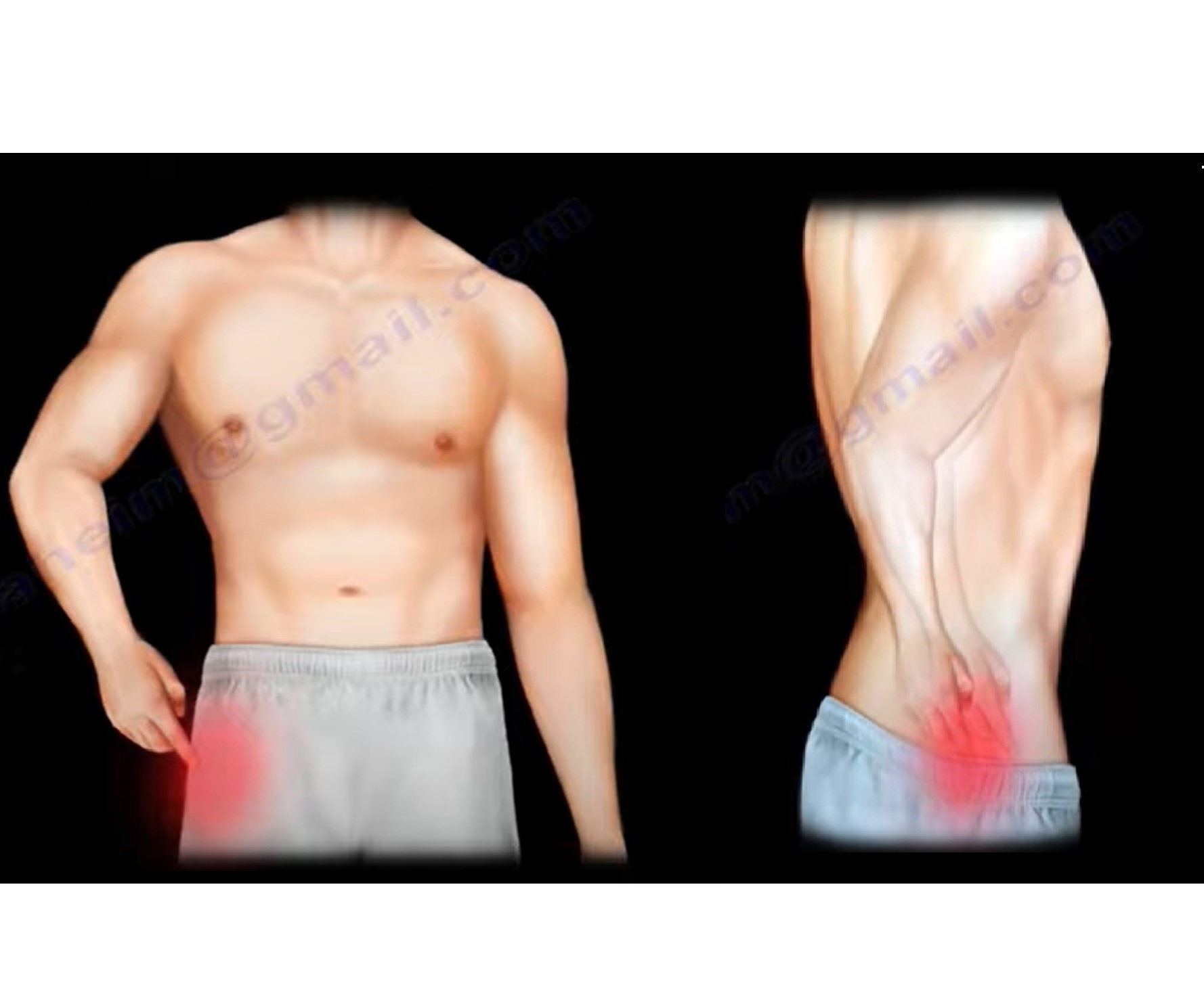
A snapping hip is a clicking or snapping sensation that occurs during hip movement.
It may be:
- Painful
- Painless
There are three main types:
- Internal
- External
- Intra-articular
2. Internal Snapping Hip
Cause
The iliopsoas tendon snaps over bony structures around the anterior hip.
It may snap over:
- Iliopectineal eminence
- Pelvic brim
The lecture also associates it with inflammation of the iliopsoas bursa, which lies between the anterior hip joint and the iliopsoas muscle or tendon.
Diagnosis
Diagnosis can be difficult.
Ultrasound or MRI may be normal, although they may demonstrate:
- Fluid within the bursa anterior to the hip joint
An iliopsoas tendon injection can be diagnostic.
Clinical Test
The diagnosis can be supported by:
- Hip abduction
- External rotation
If this reproduces the snapping or clicking, it supports an internal snapping hip.
3. External Snapping Hip
The external type is described as iliotibial band syndrome.
Cause
The iliotibial band becomes thickened and snaps over the:
Greater trochanter
Associated problems may include:
- Bursitis
- Adhesions
- Gluteus maximus inflammation
- Edema
Clinical Examination
Direct palpation over the greater trochanter may reproduce the snapping.
The patient can also be positioned on the side while the examiner places the hip into extension and rotation and feels for the pop.
4. IT Band Tightness Test
The lecture describes a test for assessing iliotibial band tightness.
Position
- Patient lies on the side.
- Unaffected leg is positioned underneath.
- Examiner abducts the affected leg.
- Examiner extends the hip.
- The leg is then allowed to lower.
Positive Test
The leg cannot descend past anatomic neutral because of IT band tightness.
The lecture describes this as the knees being unable to come together.
5. Intra-articular Snapping Hip
The intra-articular type originates from pathology within the hip joint.
Important causes include:
- Labral tear
- Loose body
Clinical Features of Labral Tear
A patient may have:
- Decreased range of motion
- Painful clicking
- Mechanical symptoms within the joint
6. MRI Arthrogram for Labral Tear
The lecture describes the labrum as a black triangular structure on the MRI arthrogram.
The contrast relationship to the labrum is important.
Normal
Contrast may enter the lateral recess adjacent to the labrum.
This is described as normal and does not necessarily indicate a tear.
Labral Tear
Contrast extending medial to the black triangular labral structure indicates a tear according to the lecture.
Best Investigation
MRI arthrogram is described as the best study for diagnosing a labral tear.
Quick Comparison
| Feature | Internal | External | Intra-articular |
|---|---|---|---|
| Main structure | Iliopsoas tendon | Iliotibial band | Labrum / loose body |
| Location | Anterior hip | Greater trochanter | Within hip joint |
| Mechanism | Iliopsoas snapping | IT band snapping over greater trochanter | Intra-articular pathology |
| Key examination | Abduction + external rotation | Palpation over greater trochanter, IT band tightness test | Painful click + reduced ROM |
| Imaging | US/MRI may be normal | Mainly clinical | MRI arthrogram |
| Important associated pathology | Iliopsoas bursitis | Bursitis, soft-tissue inflammation | Labral tear, loose body |
Exam Pearls
- Internal snapping – iliopsoas tendon
- External snapping – IT band over greater trochanter
- Intra-articular snapping – labral tear or loose body
- Anterior snapping – think iliopsoas
- Lateral snapping – think IT band
- Painful click + reduced ROM – think labral pathology
- Iliopsoas injection can be diagnostic
- MRI arthrogram is the best study for labral tear
- Contrast in the lateral recess can be normal
- Contrast medial to the labral structure indicates a tear according to the lecture.

Leave a Reply