Courtesy: Rishi Mugesh Kanna, Consultant Spine Surgeon, Ganga Hospital, Coimbatore, India
Spinal Injections
-
Spinal injections are defined as injections performed in and around the vertebral column, spinal cord, and nerve roots.
-
They may involve one or more of the following agents:
-
Local anesthetics for diagnostic nerve blocks
-
Steroids for epidural steroid injections
-
Contrast dye for discography
-
-
Spinal injections may be:
-
Diagnostic
-
Therapeutic
-
Both diagnostic and therapeutic
-
Classification of Spinal Injections
Diagnostic Injections
-
Discography for evaluation of axial back pain
-
Facet joint injections
-
Diagnostic nerve root blocks
Diagnostic and Therapeutic Injections
-
Epidural steroid injections:
-
Transforaminal
-
Interlaminar
-
Caudal
-
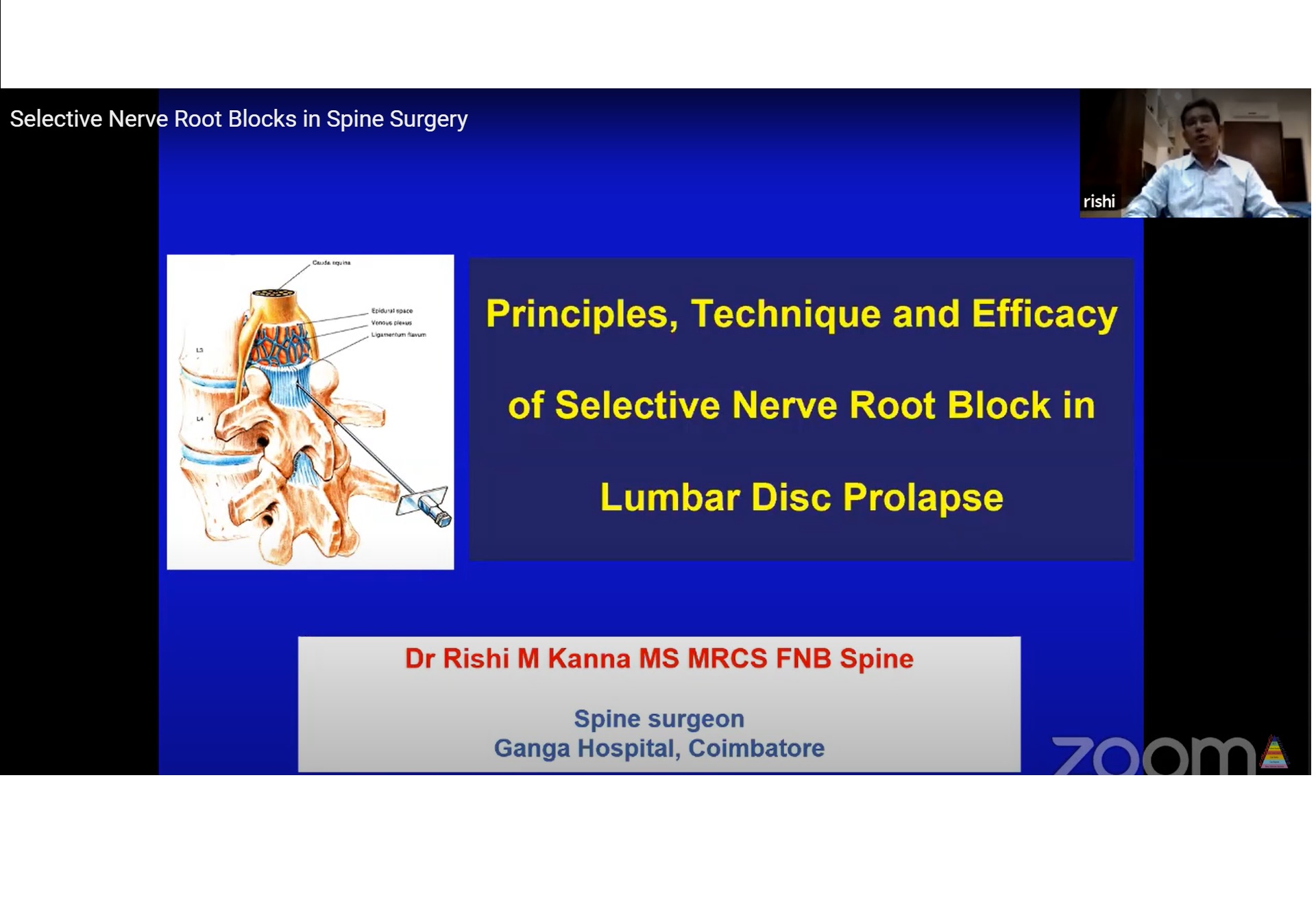
Transforaminal Epidural Steroid Injection
(Selective Nerve Root Block)
-
Used for:
-
Unilateral radiculopathy (preferred)
-
Bilateral radiculopathy
-
-
Indications include:
-
Lumbar disc prolapse
-
Lumbar canal stenosis
-
Facet joint cyst
-
Spondylolisthesis
-
Pathophysiology of Lumbar Radiculopathy
-
Radiculopathy occurs due to:
-
Mechanical compression of the nerve root
-
Chemical inflammation around the nerve root
-
-
Radicular pain is primarily produced by inflammation
-
Mechanisms include:
-
Autoimmune response to disc material in acute herniated nucleus pulposus
-
Venous congestion and ischemia in chronic disc prolapse and foraminal stenosis
-
Principle of Steroid Use
-
Steroids reduce acute inflammation around the nerve root
-
This helps break the pain cycle and provides symptom relief
-
Mechanisms of action include:
-
Suppression of nociceptive transmission in unmyelinated C fibers
-
Inhibition of phospholipase A2 and inflammatory mediators
-
Reduction of capillary permeability
-
Membrane stabilizing effect
-
Transforaminal Epidural Steroid Injection: Key Features
-
Widely accepted standard of care in many countries
-
Day-care procedure
-
Provides immediate analgesic effect
-
Cost-effective
-
Performed under local anesthesia
-
Patient positioned prone
-
Fluoroscopy guidance is essential
Needle Placement and Safe Triangle Concept
-
Needle is placed into the neuroforamen toward the safe triangle
-
Boundaries of the safe triangle:
-
Superior border: Inferior margin of the pedicle
-
Medial border: Lateral margin of the pedicle
-
Hypotenuse: Oblique line passing inferolaterally from the inferomedial corner of the pedicle
-
Steps of Transforaminal Epidural Injection
-
Obtain an oblique fluoroscopic view to identify the scotty dog appearance.
-
Infiltrate the skin and deeper tissues with local anesthetic.
-
Advance a 22 gauge spinal needle toward the safe triangle.
-
Confirm needle position on lateral view with the tip located in the neural foramen below the pedicle.
-
Inject contrast dye, such as iohexol, to confirm nerve root outline.
-
Inject the final drug mixture around the nerve root:
-
Triamcinolone 80 milligrams
-
Bupivacaine 0.5 percent, 2 milliliters
-
Post-Injection Protocol
-
Patient is mobilized within 30 minutes
-
Discharge is possible within 2 hours
-
Approximately 5 to 10 percent of patients may develop:
-
Temporary numbness
-
Mild weakness
-
-
These symptoms usually resolve within 4 hours
-
Return to normal work is permitted from the next day
-
Clinical review at 6 weeks
Selection of Nerve Root for Injection
-
Applicable from lumbar nerve roots L2 to L5
-
For posterolateral disc prolapse:
-
Target the traversing nerve root
-
Example: L5 nerve root in L4–L5 disc prolapse
-
-
For foraminal or far-lateral disc prolapse:
-
Target the exiting nerve root
-
Example: L4 nerve root in L4–L5 disc prolapse
-
-
For posterocentral disc prolapse:
-
Target the traversing nerve root on the more symptomatic side
-
Factors for Successful Transforaminal Injection
-
Accurate clinical and magnetic resonance imaging correlation
-
Correct identification of involved nerve root
-
Correlation of magnetic resonance imaging with fluoroscopy, especially in lumbosacral transitional vertebrae
-
Precise needle placement
-
Clear radiculogram
-
Reproduction of typical radicular pain during contrast injection
Frequency of Injections
-
In appropriately selected patients, success rate exceeds 75 percent
-
If pain recurs after 6 weeks, repeat injection may be considered
-
Conventionally, no more than 3 injections are recommended
-
If no relief is achieved within 1 week, or pain recurs within 6 weeks, surgical options should be discussed
Interlaminar Epidural Steroid Injection
-
Most commonly performed by anesthesiologists
-
Injection given through the interlaminar space, usually at L3–L4 or L4–L5
-
Midline approach is used
-
Loss of resistance technique employed
-
Fluoroscopy is usually not used
-
Drug spread is less targeted compared to transforaminal injection
Caudal Epidural Steroid Injection
-
Injection administered through the sacral hiatus
-
Sacral hiatus is a midline defect between the S4 and S5 laminae
-
Thecal sac typically ends at the S2 level
-
Lowest risk of accidental dural puncture
-
Drug spread is diffuse and nonspecific
Comparison of Epidural Injection Techniques
-
Transforaminal injection:
-
Highly target-specific
-
Small, concentrated drug volume
-
Very high efficacy
-
Requires greater technical skill
-
Ideal for unilateral radiculopathy
-
-
Interlaminar injection:
-
Moderately specific
-
Larger and more diluted drug volume
-
Moderate efficacy
-
Suitable for unilateral or bilateral radicular pain
-
-
Caudal injection:
-
Least specific
-
Largest and most diluted drug volume
-
Lower efficacy
-
Easiest to perform
-
Useful for bilateral or nonspecific symptoms
-
Outcomes
-
Transforaminal injections show superior outcomes compared to interlaminar and caudal techniques
-
Best suited for:
-
Acute
-
Single-level
-
Unilateral radiculopathy
-
-
Approximately 70 to 80 percent of patients show good pain relief at 2 weeks
Poor Prognostic Factors
-
Predominant sensory symptoms
-
Higher Oswestry Disability Index score
-
Non-manual occupation
-
Presence of lumbosacral transitional vertebra
-
Diffuse or splash pattern on radiculogram
Take-Home Messages
-
Spinal injections play both diagnostic and therapeutic roles
-
Radicular pain is best treated with transforaminal epidural injection
-
Selective nerve root blocks are highly specific and target-oriented
-
Best results are seen in acute and unilateral radiculopathy
-
Technique has a short learning curve, low complication rate, and good outcomes

Leave a Reply