
Courtesy: Prof Nabil Ebraheim, University of Toledo, Ohio, USA
Topic / Overview
Sciatica is defined as pain radiating along the anatomic course of the sciatic nerve, originating from the lower back, traversing the buttock and posterior thigh, and extending into the lower leg and foot.
-
Core Concept: Sciatica is a clinical symptom of an underlying condition, not a definitive diagnosis. It typically affects one side of the body.
-
Epidemiology and Natural History: Approximately 3 million individuals suffer from sciatica. It is frequently self-diagnosed and self-limiting, typically resolving spontaneously within a few weeks to a few months.
-
Terminology Distinction:
-
True Sciatica / Lumbar Radiculopathy: Arises from irritation or compression of lumbar or sacral nerve roots within the spine (e.g., disc herniation).
-
Non-Radicular Sciatica: Arises from irritation along the nerve course distal to the roots (e.g., piriformis syndrome) or referred pseudo-radicular pain (e.g., sacroiliac joint dysfunction).
-
Anatomy of the Sciatic Nerve
-
Roots of Origin: Formed by five individual roots:
-
Two lumbar roots: L4, L5
-
Three sacral roots: S1, S2, S3
-
-
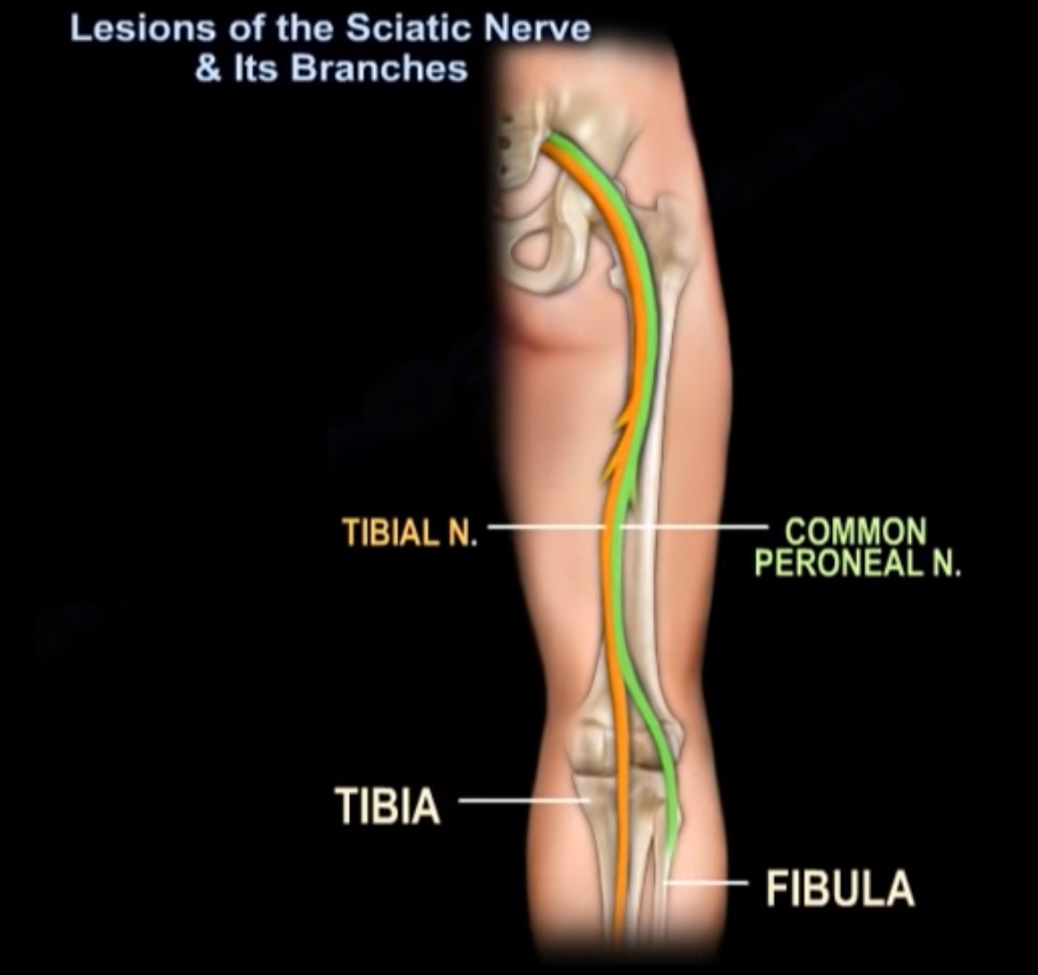
Course and Distribution: The five nerve roots combine into a single trunk in the pelvis, exit through the buttock (passing deep/inferior to the piriformis muscle), descend through the posterior thigh, and divide in the thigh and lower leg to deliver motor innervation and sensory distribution to specific muscle compartments and cutaneous territories of the leg and foot.
Etiology and Pathomechanics
The speaker outlines five primary etiologies responsible for sciatic symptoms:
1. Lumbar Disc Herniation
-
Pathomechanics: The intervertebral disc serves as an intervertebral cushion. A herniation occurs when the soft inner material (nucleus pulposus) tears through and leaks out of the stronger outer fibrous layer (annulus fibrosus), compressing and irritating adjacent nerve roots.
-
Common Pattern: Herniations most frequently occur in a posterolateral direction.
-
Predominant Level: Most commonly involves L5–S1, resulting in direct compression of the S1 nerve root.
-
Sciatica represents one of the most classic clinical manifestations of lumbar disc herniation.
2. Degenerative Disc Disease (DDD)
-
Pathomechanics: Progressive degeneration leads to secondary osteophyte formation (bone spurs) that project into the neural pathways, directly impinging on exiting or traversing nerve roots.
-
Level and Root Correlation: Involvement at the L4–L5 disc level typically compresses the L5 nerve root.
3. Isthmic Spondylolisthesis
-
Pathomechanics: Driven by a defect in the pars interarticularis (spondylolysis), allowing forward translation (-listhesis or slip) of one vertebral body over the caudal segment. The resulting slip, combined with disc space collapse and local stress fracture changes, causes mechanical nerve pinching.
-
Level and Root Correlation:
-
L5–S1 slip: Involves the L5 nerve root.
-
L4–L5 slip: Involves the L4 nerve root.
-
4. Lumbar Spinal Stenosis
-
Pathomechanics: Circumferential narrowing of the central spinal canal and neural foramina resulting from:
-
Hypertrophy and enlargement of the facet joints
-
Hypertrophy of the ligamentum flavum
-
Intervertebral disc degeneration
-
General spinal osteoarthritis
-
-
Clinical Characteristics: Neurogenic claudication characterized by leg pain, weakness, muscle cramps, burning, and a subjective “heavy sensation” in the extremities.
-
Postural Dynamics:
-
Relieving factors: Spinal flexion or leaning forward (classically described as leaning over a shopping cart).
-
Aggravating factors: Spinal extension and standing still.
-
5. Piriformis Syndrome
-
Pathomechanics: Entrapment and direct mechanical irritation of the sciatic nerve trunk as it passes beneath the piriformis muscle belly in the buttock.
-
Distinction: Represents local extra-spinal sciatic nerve compression rather than a true lumbar radiculopathy, although it clinically produces sciatica.
6. Sacroiliac (SI) Joint Dysfunction (Differential Entity)
-
Does not produce true radiculopathy, but clinical manifestations mimic both lumbar radiculopathy and piriformis syndrome. These three conditions frequently overlap and can create diagnostic confusion.
Clinical Features and Physical Examination
Clinical Semiology of Sciatic Pain
-
Quality of Pain: Sharp, shooting, electric shock-like sensation traveling down the extremity, often accompanied by burning, tingling, and paresthesias (“pins and needles”).
-
Sensory/Motor Findings: Objective numbness, subjective/objective weakness, and an inability to stand or ambulate in severe presentations. Symptoms correspond to the specific root compressed.
-
Relationship to Axial Back Pain: Patients often have concurrent low back pain, but the radiating leg pain is characteristically far more severe than the back pain.
-
Mechanical Exacerbation: Sudden maneuvers, position shifts, coughing, and sneezing increase intrathecal/mechanical pressure and aggravate pain.
-
Positional Relieving/Aggravating Factors in Discogenic Sciatica:
-
Worse: Sitting (places tension and stretch directly on the sciatic nerve trunk, causing irritation) and prolonged standing.
-
Better: Walking or lying recumbent.
-
Diagnostic Tests
-
Straight Leg Raising (SLR) Test (Tension Sign):
-
Primary physical examination maneuver for lumbar disc herniation.
-
Positive Threshold: Reproduction of radiating leg pain and paresthesia between 30° and 70° of passive hip elevation.
-
Key Distinction: The test is considered positive only when it reproduces the patient’s radicular leg pain, not isolated axial low back pain.
-
Diagnostic Utility: Highly sensitive for identifying L5 or S1 radiculopathy.
-
Diagnostic Utility in Stenosis: SLR is rarely positive in lumbar canal stenosis; neurological examination is completely normal in over 50% of stenosis patients.
-
-
Imaging Modality of Choice: Magnetic Resonance Imaging (MRI) is the definitive imaging study to identify disc herniation, neural compression, and canal morphology.
Differential Diagnosis: Neurogenic vs. Vascular Claudication
Every patient evaluated for suspected lumbar spinal stenosis must undergo an assessment for peripheral vascular disease, specifically including palpation of peripheral pulses.
-
Response to Walking: Both conditions produce symptomatic claudication upon ambulation.
-
Response to Sitting: Both conditions experience symptom resolution when seated.
-
Response to Standing Still:
-
Lumbar Stenosis (Neurogenic): Symptoms persist or worsen because the spine remains extended.
-
Vascular Claudication: Symptoms resolve promptly because metabolic demands of the calves drop.
-
-
Response to Stationary Cycling:
-
Lumbar Stenosis (Neurogenic): Relieves symptoms due to the maintained forward-flexed posture of the lumbar spine opening the spinal canal.
-
Vascular Claudication: Aggravates symptoms due to increased peripheral muscular oxygen demand.
-
Management Principles: Lumbar Disc Herniation
Non-Operative Treatment
-
Prognosis: Approximately 90% of patients improve within 1 month using conservative management.
-
Modalities: Brief rest, physiotherapy, and non-steroidal anti-inflammatory medications.
Surgical Management
-
Indications for Surgery:
-
Progressive motor weakness.
-
Persistent, disabling radicular pain lasting greater than 6 weeks refractory to non-operative measures.
-
-
Ideal Surgical Candidate Profile:
-
Severe sciatica (predominantly leg pain exceeding back pain)
-
Positive tension sign (positive straight leg raise test)
-
Concordant abnormal neurological examination findings
-
Confirmatory MRI findings precisely matching the clinical and neurological level
-
-
Surgical Techniques:
-
Hemilaminotomy
-
Discectomy
-
Outcomes and Evidence Cited
-
Early vs. Late Benefits: Surgical decompression provides significantly faster pain relief, functional recovery, and patient satisfaction compared to non-operative treatment in the short-to-intermediate term.
-
Long-Term Equivalence (4-Year Outcome): There is no significant difference in work status between surgical and non-surgical management at 4-year follow-up.
-
Postoperative Rehabilitation: Patients may resume intense physical activities at 4 to 6 weeks postoperatively.
Key Take-Home Points
-
Sciatica is an umbrella symptom, not an anatomical diagnosis; identifying the precise underlying mechanical cause (disc herniation, canal stenosis, listhesis, or piriformis compression) is required.
-
Natural history favors resolution: 90% of acute disc herniations improve within 4 weeks under conservative management.
-
A positive straight leg raise is defined by leg pain reproduction between 30° and 70°; isolated back pain does not constitute a positive tension sign.
-
Surgery achieves accelerated relief and earlier functional recovery, but 4-year return-to-work rates do not differ significantly from non-operative treatment.
-
Always palpate distal pulses in older adults presenting with ambulation-induced leg pain to distinguish spinal stenosis from vascular claudication.
Exam Pearls
-
Root Affliction Rule in Spondylolisthesis:
-
In isthmic spondylolisthesis at L5–S1, the exiting L5 root is compressed.
-
In isthmic spondylolisthesis at L4–L5, the L4 root is compressed.
-
-
Root Affliction Rule in Posterolateral Disc Herniation:
-
A posterolateral disc herniation at L5–S1 compresses the traversing S1 root.
-
A disc herniation at L4–L5 typically involves the L5 root.
-
-
The “Bicycle Test” Paradigm:
-
Relief on a stationary bike indicates neurogenic claudication from lumbar stenosis (flexion opens the canal).
-
Exacerbation on a stationary bike confirms vascular claudication (muscular ischemia overrides posture).
-
-
Four-Year Surgical Horizon:
-
While hemi-laminotomy/discectomy hastens recovery, long-term employment/work status at 4 years demonstrates parity with conservative cohorts.
-


Leave a Reply