Courtesy: Prof Nabil Ebraheim, University of Toledo, Ohio, USA
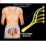
Sciatic nerve- origin, variations and course
Origin
- Sciatic nerve is the largest nerve in the body. The Sciatic nerve arises from lumbosacral plexus in the pelvis.
- Sciatic nerve plus S4 is lumbosacral plexus
- The ventral rami of L4 to S3 nerve roots unite to form the sciatic nerve
- The sciatic nerve has two components: Tibial nerve and common peroneal nerve. The tibial nerve arises from the ventral divisions of all roots in the lumbosacral plexus from L4-S3(supplies the posterior muscles of the leg).
- The common peroneal nerve arises from the dorsal division from L4-S2 and does not arise from S3(supplies the anterior muscles of the leg).The sciatic nerve ends just above the popliteal fossa by dividing into the common peroneal and tibial nerve.
Variations
- The level of division of the sciatic nerve is variable. Division of sciatic nerve usually occurs at the middle or the lower third of the thigh but in 10% of patients the tibial nerve and common peroneal nerve can be separated at the greater sciatic foramen.
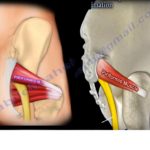
- There are 4 or more anatomic variations for the sciatic nerve in relation to the pyriformis muscle.
1.Normal relationship with the sciatic nerve -passing beneath the pyriformis muscle.
2.Piriformis divided into 2 parts with the peroneal division of the sciatic nerve passing between the 2 parts of the pyriformis muscle.
3.Peroneal division of the sciatic nerve passes over the muscle and tibial division passes beneath the undivided piriformis muscle(0.86%).
4.The entire nerve passes through the divided pyriformis muscle (0.13%).
The tibial nerve is the medial nerve and it descends down vertically towards the tibial. The common peroneal nerve is lateral, one division is tibial and the other is fibula.
Course
- The sciatic nerve leaves the pelvis through the greater sciatic foramen, normally below the piriformis muscle and anterior to it, to reach the gluteal region under cover of the gluteus maximus muscle.
- From the gluteal region where the sciatic nerve is covered by gluteal maximus ,the nerve runs downwards to the back of the thigh.
- The sciatic nerve lies between the greater trochanter and ischium and as it descends, it passes anterior to the piriformis muscle and crosses posterior to the tendon of gemellus muscle, the obturator internus and the quadratus femoris muscles.
- The sciatic nerve is anterior to the piriformis but posterior to the obturator internus. When placing the sciatic nerve retractor in the lesser sciatic notch, the obturator internus tendon is between the retractor and the nerve.
- The sciatic nerve is closely related to the posterior aspect of the acetabulum and can be injured due to dislocation or from traction or retractors of surgery. In the thigh the sciatic nerve descends on the posterior aspect of the adductor magnus muscle in a midline direction.
- In the upper aspect of the thigh,the sciatic nerve is covered by the long head of the biceps femoris muscle (nerve is deep to the biceps femoris).The nerve descends in the thigh close to the midline crossed obliquely by the long head of the biceps femoris muscle over the sciatic nerve looks somewhat like X
- In the rest of it’s course in the thigh, the sciatic nerve is covered by the biceps femoris and the hamstring muscles.
Branches
- High in the thigh before the nerve divides, the sciatic nerve supplies the hamstring muscles, which are the long head of the biceps femoris, the semimembranosus and the semitendinosus .
- It also supplies the ischial part of the adductor magnus muscle. Excluding the short head of the biceps femoris, all branches to the muscles come from the medial side of the sciatic nerve.
- The short head of the femoris is innervated by the common peroneal nerve.(The lateral border of sciatic nerve is safer during dissection or surgery.
- On its medial side, the sciatic nerve is accompanied by the posterior cutaneous nerve of the thigh and a small artery called the inferior gluteal artery.
- The sciatic nerve is close to the hamstrings tendon(about 1 cm lateral to the proximal hamstring tendons).
- The nerve should be identified, dissected away and neurolysis may be needed, especially in cases of chronic repair of the hamstring muscle(cases that require mobilization of the retracted tendons).
- These chronic cases may cause an increased difficulty in neurolysis of the sciatic nerve.

Leave a Reply