Revision ACL Reconstruction Principles
Prevalence of ACL Reconstructions
-
ACL reconstruction rate in the U.S.: 74.6 per 100,000 people
-
Positive outcomes in 75%–97% of cases
-
Higher graft rupture rates seen in patients under 20 years old
Consequences of ACL Deficiency
-
Knee instability
-
Restricted range of motion
-
Increased risk of meniscal injuries
-
Progression to degenerative joint disease
-
Arthritis development
-
Limitations in physical activity
Causes of Graft Failure
-
MARS study: Multiple causes in 37% of revision cases
-
Traumatic re-injury: Most common single cause (32%)
-
Technical errors: Account for 24%
-
Biological issues: Contribute to 7%
-
Common technical errors:
-
Misplaced tunnels (most significant)
-
Incorrect or failed hardware
-
Undetected limb malalignment
-
Tunnel Malpositioning Effects
-
Anterior femoral tunnel: Causes graft over-tension, limited flexion, potential failure
-
Vertical femoral tunnel: Preserves AP stability, reduces rotational control
-
Tibial tunnel issues:
-
Anterior placement: Impingement during extension
-
Posterior placement: Graft laxity in flexion, possible PCL impingement
-
Additional Risk Factors for Graft Failure
-
Fixation issues:
-
Critical to maintain graft tension
-
Bone-tendon-bone: Can fail at bone interface or screw pull-out
-
Soft-tissue: Susceptible to fastener misplacement or tension problems
-
-
Unaddressed issues during primary surgery:
-
Posterior tibial slope >12° or varus alignment = increased graft stress
-
Missed injuries (posterolateral/posteromedial corners, medial meniscus) raise failure risk
-
Types of Traumatic Graft Failure
-
Early-stage: Before graft incorporation
-
Late-stage: Post-return to activity
-
Early ROM-focused physiotherapy is encouraged
-
Avoid aggressive strengthening exercises
-
Osteolysis >15 mm requires bone grafting prior to revision
Preoperative Considerations
-
Evaluate need for staged vs. concomitant procedures
-
Address limb alignment to avoid graft stress
-
Corrective procedures:
-
Posterior tibial slope: Anterior closing wedge osteotomy
-
Varus: High tibial osteotomy (HTO)
-
Valgus: Lateral opening wedge distal femoral osteotomy or medial closing wedge DFO
-
Single-Stage vs. Two-Stage Revision
-
Decision depends on:
-
Tunnel integrity and placement options
-
Presence of tunnel osteolysis or conflict
-
-
Single-stage indications:
-
Tunnel osteolysis <15 mm
-
No severe malalignment or motion deficits
-
Resolved infections
-
-
Motion deficits to address:
-
20° flexion loss or >5° extension loss must be treated pre-revision
-
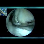
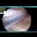
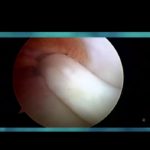
Surgical Technique Overview
-
Initial Arthroscopy:
-
Assess tunnel positioning and joint condition
-
Check for cartilage damage, meniscal tears, loose bodies
-
Single-Stage Revision Procedure
-
Debridement of previous graft
-
Expose tunnels clearly
-
Remove interfering hardware only
-
Tunnel overlap not a contraindication—requires stable fixation strategy
-
Options for tunnel management:
-
Bone graft or substitute
-
Immediate re-drilling
-
-
Avoid stacking interference screws
-
If fixation is compromised, convert to two-stage revision
Two-Stage Revision Procedure
-
Purpose: Restore bone stock and prepare for new tunnels
-
Remove all hardware
-
Ream and graft tunnels with allograft (chips or dowels)
-
Dowels rehydrated with sterile saline; matched to reamer size
-
Wait 3–6 months for bone integration
-
Confirm integration with X-ray or CT
Graft & Fixation Choices
-
Autograft: Lower failure rates, better outcomes per some reviews
-
Harvesting options:
-
Prefer ipsilateral
-
Use contralateral if ipsilateral is not viable
-
-
Fixation:
-
Use interference screws when bone stock allows
-
Suspensory/extracortical fixation as alternatives
-
Extraarticular Supplementation
-
ALC (anterolateral complex) critical for rotational control
-
Injuries to ALC common with ACL tears
-
Augmentation options:
-
LET or anterolateral ligament reconstruction
-
Useful in:
-
Patients <25 years
-
Multiple revisions
-
High-risk rotational sports
-
-
-
Preferred technique: Modified Lemaire (iliotibial band tenodesis)
-
Caution: Risk of over-constraining the joint
Postoperative Rehabilitation
-
Weight-bearing:
-
Immediate if isolated ACL revision
-
Delayed if combined with osteotomy/meniscal/cartilage procedures
-
-
Early rehab:
-
Heel slides, quadriceps sets, ROM exercises
-
-
Strength phase (~6 weeks):
-
Closed chain exercises to build muscle safely
-
-
Return to Sports:
-
Expected in 9–12 months
-
Based on strength, tolerance, and overall recovery
-
Outcomes
-
Generally inferior to primary ACL reconstructions
-
Grassi et al: Revision ACLs scored 7.8 points lower (Lysholm scale)
-
Mohan et al: 6% failure rate in revision ACLs
Future Directions
-
Improved tunnel management
-
Fast-setting bone graft substitutes
-
Accelerated second-stage procedures
-
Expanded use of LET in high-risk cases
-
Posterior tibial slope correction gaining recognition as key to reducing graft failure risk
Leave a Reply