Introduction & Early Designs
-
1974: Concept introduced by Charles Neer
-
Intended to treat:
-
Glenohumeral arthrosis
-
Rotator cuff deficiency
-
-
-
Early implant designs faced major limitations:
-
Glenoid component loosening
-
Implant breakage due to constrained designs
-
Lateralized center of rotation leading to high mechanical stress
-
Grammont’s Contribution (1985)
Introduced a reverse ball-and-socket design based on four key principles:
-
Medialization of center of rotation
-
Reduces mechanical torque at the glenoid–implant interface
-
-
Inferior positioning of humerus
-
Increases deltoid tension
-
Improves deltoid recruitment
-
-
Fixed center of rotation
-
Enhances implant stability
-
-
Large glenosphere
-
Increases range of motion
-
Allows a semi-constrained articulation
-
These principles form the foundation of modern RTSA.
What is Reverse Shoulder Arthroplasty?
-
A form of shoulder arthroplasty where:
-
Convex glenosphere is placed on the glenoid
-
Concave cup is placed on the humeral side
-
-
Reverses normal shoulder anatomy:
-
Glenoid fossa ? baseplate + glenosphere
-
Humeral head ? stem + concave polyethylene cup
-
-
Alters shoulder biomechanics by:
-
Medializing and inferiorly shifting the center of rotation
-
Increasing deltoid tension and moment arm
-
-
Result:
-
Improved shoulder elevation despite rotator cuff deficiency
-
Indications for Reverse Shoulder Arthroplasty
-
Cuff tear arthropathy
-
Pseudoparalysis due to massive rotator cuff tear (without arthritis)
-
Multiple failed rotator cuff repairs with:
-
Poor function
-
Anterosuperior instability
-
-
Three- and four-part proximal humerus fractures in elderly patients
-
Proximal humeral nonunions
-
Greater tuberosity malunions
-
Failed hemiarthroplasty with anterosuperior escape
-
Tumour-related shoulder pathology
Patient Selection Criteria
-
Essential requirements:
-
Intact and functioning deltoid muscle
-
Adequate glenoid bone stock
-
No active infection or sepsis
-
-
Avoid in patients with:
-
Severe neurological disorders (Parkinson’s disease, Charcot joint, syringomyelia)
-
Very high functional demands unsuitable for RTSA
-
Biomechanics of RTSA
-
Medialized and inferior center of rotation:
-
Allows deltoid to act on a longer lever arm
-
Compensates for deficient rotator cuff
-
-
Benefits:
-
Improved shoulder abduction and elevation
-
-
Limitations:
-
Limited improvement in internal and external rotation
-
-
External rotation may be improved with:
-
Latissimus dorsi transfer in selected cases
-
Preoperative Planning
Radiographs
Recommended views:
-
True AP (Grashey view)
-
Assess arthritis
-
Identify superior humeral migration
-
-
Axillary lateral view
-
Detect posterior glenoid wear
-
-
Scapular Y view
-
Evaluate scapular alignment
-
CT Scan
Indicated when axillary view is inadequate or deformity is suspected:
-
Evaluates:
-
Glenoid version
-
Glenoid bone stock
-
Bone quality and osteopenia
-
-
Helps plan:
-
Glenoid correction
-
Implant positioning
-
MRI
Used selectively to:
-
Assess rotator cuff integrity
-
Evaluate fatty infiltration
-
Plan soft-tissue management in complex cases
Contraindications to RTSA
Absolute
-
Global deltoid deficiency
-
Active infection
-
Severe glenoid bone loss preventing fixation
Relative
-
Partial deltoid deficiency
-
Axillary nerve dysfunction
-
Severe glenoid osteoporosis
-
Surgeon inexperience
-
Younger age (<70 years) – now acceptable in select cases
Surgical Approaches for RTSA
Deltopectoral Approach (Preferred)
Advantages:
-
Preserves deltoid muscle
-
Lower risk of axillary nerve injury
-
Better exposure of inferior glenoid
-
Allows extension inferiorly if required
-
Enables simultaneous latissimus dorsi transfer
Disadvantages:
-
Requires subscapularis takedown
-
Extensive capsular release needed
-
Limited posterior glenoid exposure
-
Risk of postoperative stiffness
Other Approaches (Not Recommended Routinely)
-
Anterolateral / transdeltoid lateral approaches
-
Higher risk of:
-
Axillary nerve injury
-
Deltoid damage
-
Structures to Be Protected
-
Cephalic vein
-
Anterior circumflex humeral artery
-
Ascending arcuate branch
-
Posterior circumflex humeral artery
-
Musculocutaneous nerve
-
Axillary nerve
-
Suprascapular nerve and artery
Patient Positioning
-
Beach-chair position
-
Trunk elevated 30–70°
-
Allows excellent visualization and access
-
Surgical Landmarks
-
Coracoid process
-
Acromion
-
Proximal humeral shaft
Incision & Exposure (Deltopectoral)
-
12–14 cm incision between:
-
Coracoid
-
Proximal humeral shaft
-
-
Develop deltopectoral interval
-
Incise clavipectoral fascia
-
Identify:
-
Conjoint tendon
-
Subscapularis tendon (divided vertically)
-
Glenoid Preparation
-
Complete soft-tissue and labral release
-
Remove biceps remnants
-
Place guide plate aligned with inferior glenoid circle
-
Central K-wire placement confirmed with pre-op planning
-
Ream glenoid surface
-
Drill central peg
-
Fix baseplate with screws
-
Implant glenosphere:
-
Larger than AP glenoid diameter
-
Slight inferior overhang to reduce scapular notching
-
Humeral Preparation
-
Humeral head osteotomy:
-
0–30° retroversion (commonly 20°)
-
Increased retroversion may improve external rotation
-
-
Canal preparation with sequential reaming
-
Trial stem insertion:
-
Recommended retroversion: ~10°
-
-
Reduce trial prosthesis and assess:
-
Stability
-
Deltoid tension
-
Impingement
-
Final Implantation
-
Cemented humeral stem fixation
-
Insert definitive polyethylene inlay
-
Reduce prosthesis
-
Tuberosity fixation:
-
Secure subscapularis and infraspinatus
-
Tie sutures to prevent superior migration
-
-
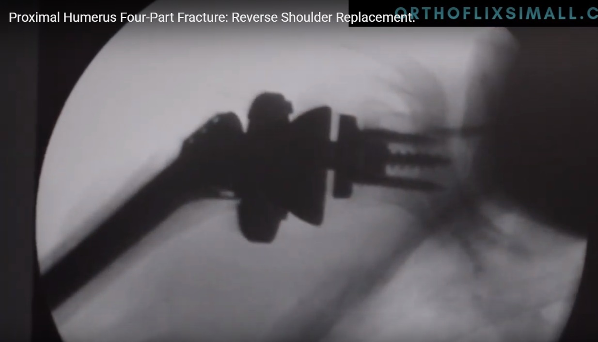
Confirm implant position with fluoroscopy
Rehabilitation Protocol
Weeks 0–2
-
Sling for comfort
-
Pendulum exercises
-
Hand, wrist, elbow motion
-
Active-assisted elevation to 90°
-
External rotation limited to ~10°
Weeks 2–6
-
Discontinue sling
-
Active-assisted ? active ROM
-
Scapular stabilization exercises
Weeks 6–12
-
Begin deltoid strengthening
-
Continue ROM
-
Avoid free weights
After 12 Weeks
-
Gradual lifting and functional use as tolerated
Outcomes
-
Better outcomes in:
-
Primary RTSA
-
-
Inferior outcomes in:
-
Post-traumatic cases
-
Revision surgery
-
-
Typical protocol:
-
Immobilization: 6 weeks
-
Active ROM: 6 weeks
-
Deltoid strengthening: 12 weeks
-
Complications of RTSA
-
Scapular notching
-
Dislocation
-
Glenoid loosening
-
Deep infection
-
Acromial or scapular spine fractures
-
Axillary nerve neurapraxia
-
Aseptic loosening of glenoid screws
Key Take-Home Messages
-
RTSA is a biomechanically driven solution for cuff-deficient shoulders
-
Proper patient selection and planning are critical
-
Deltoid integrity is essential
-
Glenoid positioning determines long-term success
-
Rehabilitation is as important as surgery
Leave a Reply