
Courtesy: Prof Nabil Ebraheim, University of Toledo, Ohio, USA
Rectus Femoris Strain: Clinical Anatomy and Diagnostic Approach
Core Takeaways
-
Overlooked Cause: Rectus femoris strains and tendinopathies are frequently underdiagnosed sources of groin and anterior hip pain in athletes.
-
Dual-Action Vulnerability: Because it is the only bi-articular quadriceps muscle (crossing both the hip and knee), it is prone to eccentric stretch and forceful contraction injuries.
-
Presentation Bifurcation: Acute strains typically present with distal or mid-thigh pain, whereas chronic overuse tendinitis centers proximally near the pelvic origin.
Functional Anatomy
-
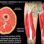
Muscle Group: Forms the superficial anterior component of the quadriceps femoris complex.
-
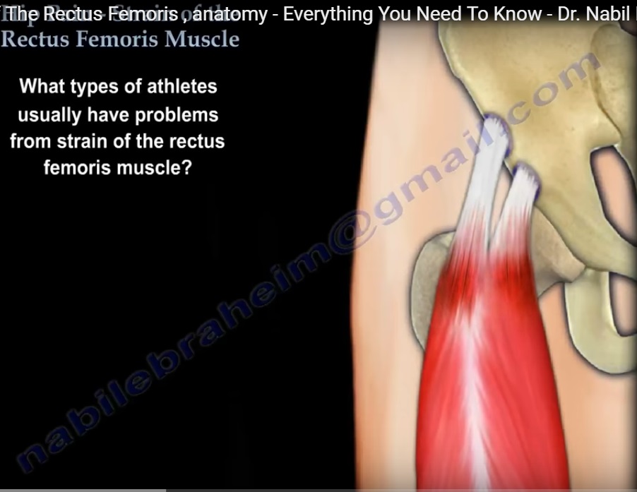
Dual Origins (Proximal Attachments):
-
Straight Head: Originates directly from the Anterior Inferior Iliac Spine (AIIS) of the pelvis.
-
Reflected Head: Originates from a groove just superior to the acetabulum.
-
-
Distal Insertion: Inserts into the superior border of the patella via the common quadriceps tendon, continuing via the patellar ligament onto the tibial tuberosity.
-
Innervation: Femoral nerve via roots L2, L3, and L4.
-
Dual Biomechanical Action:
-
Flexes the thigh at the hip joint.
-
Extends the leg at the knee joint.
-
Mechanism of Injury & High-Risk Athletes
-
At-Risk Sports: Common in soccer players, sprinters/runners, baseball players, and hockey players.
-
Acute Traumatic Onset:
-
Forceful eccentric contraction (e.g., violent hip hyperextension with knee flexion during a soccer kick or explosive acceleration from a standing start).
-
Causes mechanical tearing of muscle fibers, typically manifesting distally along the mid-to-lower muscle belly or near the knee.
-
-
Chronic Overuse (Tendinopathy):
-
Repetitive tensile overload leading to micro-tearing, scar formation, adhesions, and collagen degeneration.
-
Behaves similarly to Achilles tendinitis or lateral epicondylitis (“tennis elbow”).
-
Pain localizes proximally near the AIIS origin.
-
-
Adolescent Population (Apophysitis / Avulsion):
-
Open, unossified apophyses are weaker than the tendon itself.
-
Sudden violent traction causes an AIIS avulsion fracture rather than an isolated mid-substance muscle tear.
-
Differential Diagnosis of Anterior Hip Pain
-
Intra-Articular Etiologies:
-
Acetabular labral tears.
-
Intra-articular loose bodies.
-
Avascular necrosis (AVN) of the femoral head.
-
Hip osteoarthritis.
-
-
Extra-Articular Etiologies:
-
Iliopsoas bursitis / tendinitis.
-
Adductor strain (“sports hernia” / classic pulled groin).
-
Tensor fasciae latae (TFL) or Iliotibial (IT) band strain.
-
Rectus femoris strain/tendinitis.
-
Physical Examination & Clinical Provocation
-
Systematic Palpation:
-
ASIS (Anterior Superior Iliac Spine): Checked to rule out sartorius origin pathology.
-
Soft Groove: Palpated immediately distal/medial to ASIS before reaching the AIIS.
-
AIIS (Anterior Inferior Iliac Spine): Targeted palpation over the straight head origin; focal tenderness indicates proximal tendinopathy or avulsion.
-
Full Muscle Belly: Palpation along the mid-thigh to distal quadriceps to detect acute localized tears or focal defects.
-
Lateral Structures: Palpate the TFL and IT band to exclude lateral tract involvement.
-
-
Provocative Maneuvers:
-
Resisted Hip Flexion: Reproduces sharp pain and reveals weakness specific to rectus femoris pathology.
-
Differentiation from Adductor Strain: Adductor injuries reproduce pain on resisted leg adduction (Compression Adduction Test), distinguishing them from isolated rectus femoris involvement.
-
Diagnostic Imaging
-
Plain Radiography (X-ray):
-
Typically negative or normal in adult soft-tissue strains.
-
Mandatory in adolescents to visualize or rule out bony AIIS avulsion fractures.
-
-
Magnetic Resonance Imaging (MRI):
-
Reserved for chronic, recalcitrant cases or when distinguishing deep intra-articular pathology from refractory high-grade tendon tears.
-
Treatment & Rehabilitation Protocol
-
Conservative Non-Operative Care (Primary):
-
Phase 1 (Protection & Symptom Control): Rest, crutches (for non-weight-bearing in avulsions or severe antalgic gait), cryotherapy (ice), and NSAIDs.
-
Phase 2 (Restoration): Structured physical therapy focused on targeted stretching, myofascial release, and progressive eccentric/concentric strengthening.
-
Phase 3 (Manual Therapy): Active Release Technique (ART) to reduce scar tissue adhesions and restore muscle glide.
-
-
Injections: Considered in non-responsive chronic tendinopathy for localized anti-inflammatory benefit.
-
Surgical Intervention: Extremely rare; limited to severe, retracted bony avulsions or failure of prolonged exhaustive conservative therapy.
Leave a Reply