Introduction
-
Foot deformities across syndromic conditions often share common underlying factors:
-
Generalized joint laxity
-
Bone deformities
-
Hypotonia and hypermobility
-
-
These deformities account for a significant proportion of orthopaedic complaints in patients with genetic and connective tissue disorders.
-
The most common contributors include:
-
Increased ligamentous laxity
-
Muscle hypotonia
-
Excessive joint mobility
-
Syndromes Covered in This Review
-
Down syndrome (DS)
-
Marfan syndrome (MFS)
-
Ehlers–Danlos syndrome (EDS)
-
Larsen syndrome (LS)
-
Osteogenesis imperfecta (OI)
Down Syndrome
General Features
-
Caused by trisomy 21 due to chromosomal nondisjunction
-
Associated musculoskeletal findings:
-
Cervical spine instability
-
Scoliosis
-
Hip and patellar instability
-
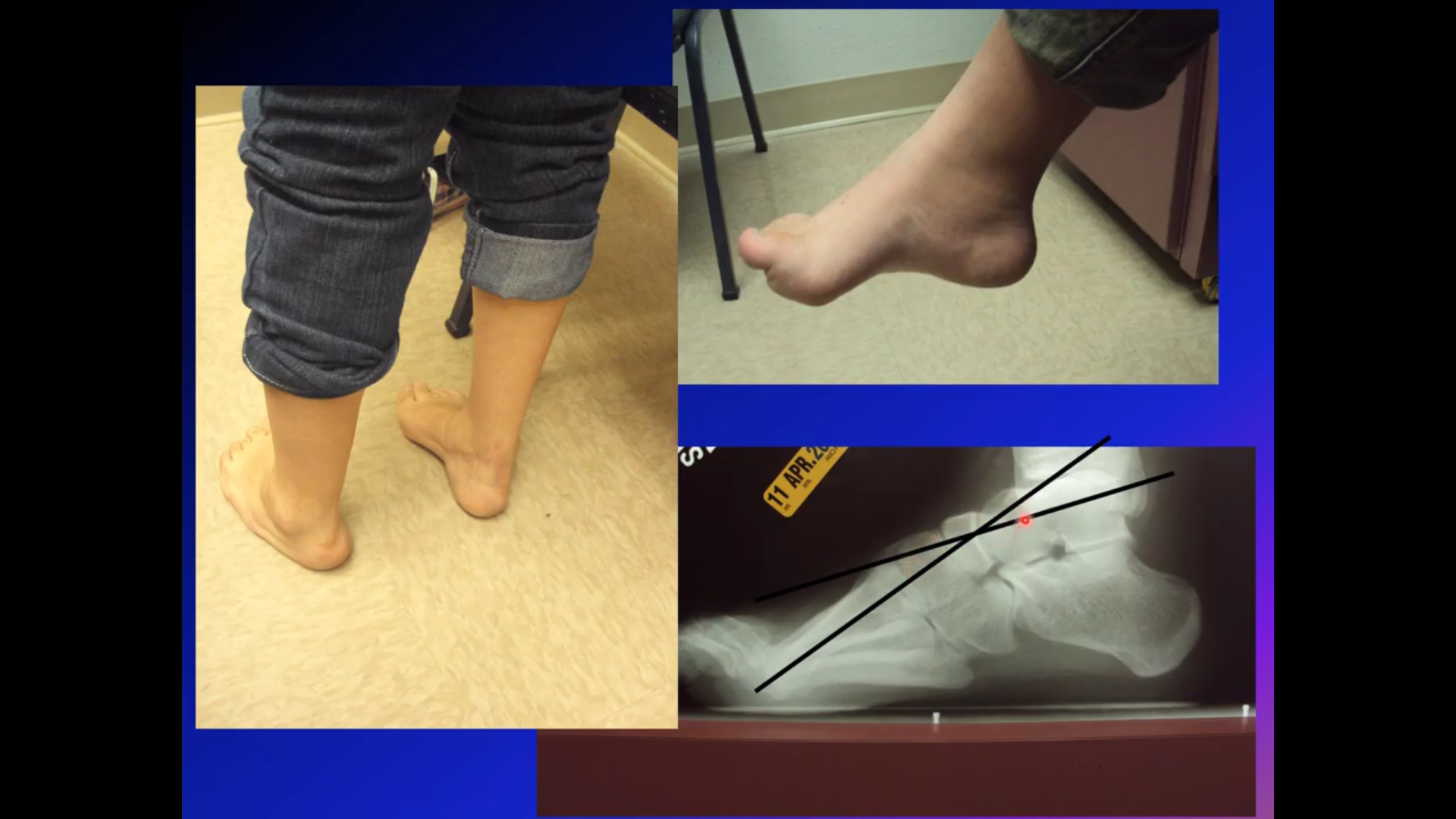
Foot and Ankle Manifestations
-
Foot deformities seen in approximately 30% of patients
-
Common deformities:
-
Pes planus
-
Hallux valgus (secondary to ligament laxity)
-
Syndactyly
-
Calcaneal valgus
-
Bony deformities
-
Management
Conservative
-
Orthotic management to improve gait mechanics and kinematics:
-
UCBL (University of California–Berkeley Lab) orthoses
-
Supramalleolar orthoses
-
Ankle–foot orthoses (AFOs)
-
-
Adjunctive physical therapy enhances outcomes
Surgical
-
Reserved for severe deformities or functional limitation:
-
First-ray realignment for severe hallux valgus
-
Exostectomy for symptomatic medial eminence
-
Marfan Syndrome
Genetic and Systemic Features
-
Autosomal dominant connective tissue disorder
-
Genetic mutations:
-
FBN1 gene (Type 1)
-
TGFBR1 and TGFBR2 genes (Type 2)
-
Multisystem Involvement
-
Ocular: lens subluxation, myopia, retinal detachment
-
Cardiovascular: aortic root dilation, aneurysm, rupture, mitral valve prolapse
-
Skeletal:
-
Scoliosis
-
Thoracic lordosis
-
Pectus excavatum
-
Hip instability
-
Joint laxity
-
Acetabular protrusion
-
Reduced bone mineral density
-
Foot Deformities
-
Predominantly:
-
Pes planus
-
Hallux valgus
-
Management
Conservative
-
Custom foot orthoses with medial longitudinal arch support
Surgical
-
Triple arthrodesis may be considered for:
-
Painful, rigid pes plano-valgus deformities
-
Improved stability and function
-
Ehlers–Danlos Syndrome
Key Characteristics
-
Heterogeneous connective tissue disorder
-
Molecular defects involving collagen types I, III, and V
Classic Triad
-
Skin hyperextensibility
-
Joint hypermobility
-
Tissue fragility
Foot and Ankle Manifestations
-
Pes planus
-
Hallux valgus with bunions
-
Hammertoes and claw toes
-
Clubfoot
-
Achilles tendon contracture
-
MTP joint subluxations
-
Chronic ankle instability leading to recurrent falls
Management
Conservative
-
Ankle bracing (e.g., ankle-stabilizing orthoses)
-
Foot orthoses to reduce pain and fatigue
Surgical
-
Often challenging due to:
-
Severe joint hypermobility
-
Poor tissue quality
-
-
Surgery reserved for selected cases with significant disability
Larsen Syndrome
Genetic Background
-
Can be autosomal dominant or recessive
-
Caused by mutations in the FLNB gene (chromosome 3p14)
Clinical Features
-
Congenital dislocations of:
-
Hips
-
Knees
-
Elbows
-
-
Spinal deformities:
-
Scoliosis
-
Cervical kyphosis
-
-
Skeletal and other features:
-
Short, wide distal phalanges
-
Craniofacial anomalies
-
Hearing loss due to malformed ear ossicles
-
Foot Deformities
-
Clubfoot deformities:
-
Equinovarus
-
Equinovalgus
-
Management
Conservative
-
Ponseti method:
-
Serial casting
-
Percutaneous Achilles tenotomy
-
Surgical
-
Reserved for resistant or recurrent cases:
-
Posteromedial soft-tissue release
-
-
High risk of recurrence despite conservative treatment
Osteogenesis Imperfecta
Pathophysiology
-
Quantitative and qualitative defects in collagen synthesis
Systemic Features
-
Short stature
-
Hearing loss
-
Blue sclera
-
Cardiopulmonary anomalies
-
Dentinogenesis imperfecta
Orthopaedic Manifestations
-
Recurrent fractures
-
Long-bone bowing
-
Genu valgum
-
Coxa vara
-
Joint laxity
-
Scoliosis
-
Skeletal fragility
Foot Deformities
-
Pes planovalgus (24–75%)
-
Skew foot (midfoot ligament laxity)
-
Clubfoot deformities
Management
Conservative
-
AFOs to prevent equinus contractures
Surgical
-
Reserved for severe deformities or failure of conservative care:
-
Osseous and soft-tissue procedures
-
Bilateral subtalar arthroereisis for pes planus
-
Subtalar and navicular–cuneiform arthrodesis for skew foot
-
Ponseti method for clubfoot, with surgery for resistant cases
-
Surgical Challenges
-
Poor bone quality may lead to:
-
Nonunion at osteotomy sites
-
Unsatisfactory outcomes after subtalar extra-articular arthrodesis
-
Key Takeaway
Foot deformities in syndromic conditions are multifactorial and progressive, requiring:
-
Early recognition
-
Individualized orthotic management
-
Careful patient selection for surgery due to high complication and recurrence rates
please provide complete description of various approaches to posterolat aspect of proximal tibia