
Courtesy: Prof Michael Hantes, President, ESSKA
Management of Post-Meniscectomy Syndrome
Introduction
Post-meniscectomy syndrome refers to pain occurring in a previously meniscectomized knee, involving either the medial or lateral compartment.
- Symptoms may develop within 1–5 years following meniscectomy, although the interval can be longer.
- Pain is the characteristic presenting symptom.
- The syndrome is primarily related to overload resulting from loss of meniscal tissue.
- The severity of symptoms is likely related to the amount of meniscal tissue removed.
- Lateral meniscectomy is particularly associated with early pain and functional problems.
Biomechanical Consequences of Meniscal Loss
The menisci play an important role in load distribution within the knee.
- The lateral meniscus is estimated to carry approximately 70% of the load in the lateral compartment.
- The medial meniscus carries approximately 50% of the load in the medial compartment.
- The peripheral tibial cartilage and subchondral bone, particularly in the lateral compartment, are considerably thinner than the central region.
- When the meniscus is removed, the previously protected peripheral cartilage is exposed to increased:
- Contact stresses
- Load
- Shear forces
Clinical Example
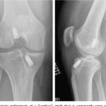
A young 18-year-old soccer player underwent subtotal lateral meniscectomy at the age of 16.
Two years later:
- Almost complete absence of the lateral meniscus was observed.
- Arthroscopy demonstrated cartilage lesions involving both the tibial and femoral surfaces of the lateral compartment.
- This illustrates the potentially rapid progression of cartilage damage following substantial lateral meniscal loss in a young patient.
Principles of Management
The primary goal in post-meniscectomy syndrome is to address the consequences of meniscal tissue loss.
Two principal options for meniscal replacement are discussed:
- Meniscal allograft transplantation
- Primarily for total or subtotal meniscal loss.
- Meniscal scaffolds
- Primarily for partial meniscal loss.
Avoid Further Meniscectomy
A further meniscectomy should not be considered simply because a patient develops pain following previous meniscectomy.
- Additional removal of meniscal tissue may further increase compartmental overload.
- A second meniscectomy is unlikely to address the underlying cause of post-meniscectomy pain.
- Progressive cartilage damage may occur following further meniscal tissue loss.
Treatment Priorities
Management should not focus solely on the meniscal defect.
The following factors should be assessed and addressed in sequence:
- Lower-limb alignment
- Ligament stability
- Meniscal deficiency
- Articular cartilage status
Key Principle
Meniscal transplantation should not be performed in an unstable knee or a malaligned knee without first addressing the instability or mechanical axis.
Meniscal Scaffolds
Meniscal scaffolds may be considered in patients with partial meniscal loss.
Collagen Meniscus Implant (CMI)
The collagen meniscus implant was among the first meniscal scaffolds introduced into clinical practice.
- It is a bovine collagen-based scaffold.
- Long-term follow-up extending beyond 20 years has been reported.
- Limitations include:
- Relatively rapid degradation
- Bovine origin
- Handling difficulties during implantation
Actifit Meniscal Scaffold
Another scaffold discussed is the Actifit meniscal scaffold.
It consists of a polymer containing:
- Approximately 80% polycaprolactone segments
- Approximately 20% more rigid polyurethane segments
A major difference from the collagen scaffold is the slower degradation process.
- Polycaprolactone hydrolysis may occur over approximately five years.
- This provides time for tissue ingrowth and gradual replacement of the scaffold.
The scaffold is available in configurations for both:
- Medial meniscal defects
- Lateral meniscal defects
Concept of Scaffold Regeneration
The scaffold is intended to act as a template for tissue regeneration.
The proposed process involves:
- Cellular ingrowth from surrounding host tissue
- Gradual degradation of the scaffold
- Replacement by meniscus-like tissue
Indications for Meniscal Scaffolding
Potential indications include:
- Partial medial or lateral meniscal loss
- Intact anterior and posterior meniscal horns
- An adequate peripheral meniscal rim
- Absence of advanced cartilage damage
The presentation suggests avoiding the procedure in:
- Patients younger than approximately 16 years
- Patients older than approximately 50 years
Patient selection should, however, be individualized.
Surgical Technique for Meniscal Scaffold Implantation
Meniscal scaffold implantation can be performed arthroscopically.
Basic Steps
- Standard anterolateral and anteromedial portals are established.
- The remaining meniscal tissue is trimmed.
- The meniscal defect is measured.
- The scaffold is prepared according to the measured defect.
- The scaffold is inserted into the joint.
- The scaffold is positioned within the meniscal defect.
- The scaffold is secured to the remaining meniscus.
Fixation
- Inside-out sutures can be used for the body and anterior portion of the meniscus.
- All-inside devices can be used for fixation of the posterior horn.
- Multiple fixation points may be used to achieve stable incorporation.
The speaker recommends slightly oversizing the scaffold to optimize its fit within the defect.
Clinical Example: Meniscal Scaffold
A 25-year-old soccer player with a previous lateral meniscectomy presented with a large lateral meniscal defect.
- Alignment was normal.
- The anterior and posterior meniscal rims were preserved.
- The defect was treated with a meniscal scaffold.
- Chondroplasty was also performed.
At approximately 12 months:
- MRI demonstrated the implanted scaffold.
- Second-look arthroscopy demonstrated the scaffold in position.
- The condition of the lateral compartment was improved compared with the preoperative appearance.
- The cartilage lesions appeared partially improved.
Outcomes of Meniscal Scaffolds
Clinical outcomes following meniscal scaffold implantation demonstrate:
- Significant reduction in pain
- Improvement in knee function
- Improvement in activity levels
- Greatest clinical improvement during the first two postoperative years
Limitations
Despite clinical improvement:
- Reported failure rates remain approximately 5–25%.
- MRI appearances are often less encouraging than clinical outcomes.
- Meniscal extrusion may be observed.
- The implant may demonstrate intermediate signal intensity.
- Partial or substantial implant absorption may occur.
- The MRI appearance does not necessarily demonstrate complete regeneration of normal meniscus-like tissue.
Long-term follow-up from the speaker’s department demonstrated significant improvements in clinical outcome scores, although MRI-based assessment showed variable scaffold maturation and morphology.
Meniscal Allograft Transplantation
Meniscal allograft transplantation has been performed clinically since the early 1980s and is therefore no longer considered an experimental procedure.
The key question is patient selection.
Indications
Potential indications include:
- Unicompartmental knee pain associated with total or subtotal meniscectomy
- Meniscal deficiency in a symptomatic young patient
- Concomitant procedure during revision ACL reconstruction in a previously meniscectomized knee
- Meniscal transplantation combined with articular cartilage restoration in a meniscus-deficient compartment
Meniscal Transplantation in Asymptomatic Patients
Routine meniscal transplantation in an asymptomatic meniscectomized knee remains controversial.
For example, in an older asymptomatic patient with previous lateral meniscectomy:
- Most experienced surgeons would not routinely recommend meniscal transplantation.
- Some surgeons may consider transplantation in selected patients with lateral meniscal deficiency.
The procedure should therefore primarily be considered for symptomatic patients, rather than simply treating an imaging finding.
Patient Selection for Meniscal Transplantation
Important factors include:
Age
Meniscal transplantation is generally considered in younger, active patients.
A precise upper age limit remains debatable, with approximately 45–55 years discussed as a practical range.
BMI
A BMI below approximately 30 kg/m² is generally preferred.
Patient Expectations
Preoperative counselling is essential.
Meniscal transplantation should not necessarily be presented as a procedure that will reliably return the patient to high-level sports.
The principal goals are:
- Pain relief
- Improved knee function
- Improved quality of life
- Preservation of the knee joint
Importance of Articular Cartilage
The condition of the articular cartilage is an important predictor of outcome.
Patients with significant cartilage damage have less favorable results following meniscal transplantation.
The presentation highlights substantially better reported survival in patients with low-grade cartilage lesions compared with those with severe cartilage damage.
Therefore:
Meniscal transplantation is best considered in appropriately selected patients with preserved or only mildly damaged articular cartilage.
Alignment and Meniscal Transplantation
Mechanical axis alignment must be assessed before transplantation.
Principle
If significant malalignment is present, realignment osteotomy should be considered.
Meniscal transplantation in a malaligned knee without correcting the mechanical axis may expose the graft to continued abnormal loading.
Meniscal Allograft Preparation and Storage
Several graft preservation methods have been described:
- Fresh viable grafts
- Fresh-frozen grafts
- Cryopreserved grafts
- Lyophilized grafts
The speaker notes that fresh-frozen grafts are currently the preferred option in his practice.
Meniscal Graft Sizing
Accurate graft sizing is important.
Radiographic Sizing
The Pollard method can be used with radiographs to estimate meniscal dimensions.
- Measurements include meniscal width and length.
- The method has a reported mean error of approximately 10%.
MRI-Based Sizing
MRI can also be used to measure the dimensions of the patient’s meniscus.
The speaker describes MRI-based sizing as his preferred approach for graft selection.
Meniscal Allograft Fixation Techniques
Several fixation techniques are available:
- Suture-only fixation
- Bone-plug fixation
- Keyhole technique
These may be performed using:
- Arthroscopic techniques
- Open techniques
- Semi-open techniques
According to the presentation, comparative studies have not demonstrated a clear difference in outcomes between the principal fixation techniques.
Surgical Technique for Meniscal Transplantation
The procedure involves several important steps.
Preparation of the Knee
- Assessment of the meniscus-deficient compartment
- Preparation of the meniscal bed
- Creation of vascular channels where appropriate
Tibial Tunnels
For a root fixation technique:
- A tunnel is created for the posterior meniscal root.
- Sutures are passed through the graft.
- An anterior tunnel is then prepared.
Care must be taken around the ACL to avoid damaging either:
- The native ACL
- An ACL graft
Graft Insertion
Graft insertion into the joint is one of the technically demanding portions of the procedure.
Once the graft is positioned:
- The posterior portion is stabilized.
- The body of the meniscus is secured using inside-out sutures.
- Multiple sutures are placed on both the tibial and femoral surfaces.
- All-inside devices can be used for the posterior portion.
The goal is stable fixation of the:
- Anterior horn
- Meniscal body
- Posterior horn
Outcomes Following Meniscal Allograft Transplantation
Meniscal transplantation can provide substantial clinical improvement.
Reported outcomes include:
- Significant pain reduction
- Improved functional scores
- Improved patient-reported outcomes
- Long-term graft survival
The presentation cites approximately 70% graft survival at 10 years, with survival decreasing over longer follow-up.
At approximately 15 years, reported survival may be in the range of 40–50%.
Return to Sports
Return to sports following meniscal transplantation is possible in selected patients.
The presentation cites:
- Approximately 92% return to play in one study involving professional and semi-professional athletes.
- More generally, reported return-to-play rates are approximately 40–70%.
Patient selection, expectations, cartilage status, associated procedures, and activity demands should all be considered.
Meniscal Transplantation Combined with ACL Reconstruction
Meniscal transplantation may be performed in conjunction with ACL reconstruction in selected patients with meniscal deficiency.
The presentation describes favorable outcomes following:
- Isolated meniscal transplantation
- Meniscal transplantation combined with ACL reconstruction and lateral extra-articular tenodesis
However, isolated meniscal transplantation may provide better outcomes than combined procedures in appropriately selected patients.
Post-Meniscectomy Arthritis and Malalignment
Progressive arthritis following meniscectomy may become a painful manifestation of the post-meniscectomy state.
In patients with:
- Meniscal deficiency
- Mechanical axis deviation
- Compartment-specific overload
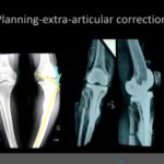
Realignment osteotomy may be required.
Important Planning Parameters
Preoperative planning should take into account:
- Overall lower-limb alignment
- Medial proximal tibial angle
- Lateral distal femoral angle
- Compartment-specific mechanical loading
High Tibial Osteotomy and Meniscal Deficiency
A key question is whether meniscal transplantation should routinely be combined with realignment osteotomy.
The presentation highlights that this remains controversial.
Medial Compartment
In patients with medial meniscal deficiency and varus alignment:
- High tibial osteotomy can provide good clinical results.
- Meniscal transplantation may not always be necessary.
- The speaker’s practice is generally to use high tibial osteotomy alone in selected medial-compartment cases.
Long-term results presented from patients with previous medial meniscectomy demonstrate good outcomes following high tibial osteotomy.
Lateral Compartment
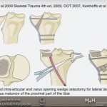
For young patients with:
- Lateral meniscal deficiency
- Valgus alignment
- Compartmental cartilage damage
A combination of:
- Distal femoral osteotomy
- Lateral meniscal transplantation
may be considered.
Key Take-Home Messages
1. Meniscal loss can produce significant compartmental overload
The menisci play an important role in load distribution. Substantial meniscal loss can lead to increased contact and shear stresses and may contribute to progressive cartilage damage.
2. Do not treat the meniscal defect in isolation
Before considering meniscal replacement, assess:
- Alignment
- Ligament stability
- Meniscal deficiency
- Cartilage status
3. Meniscal scaffolds are an option for partial meniscal loss
Scaffolds can provide:
- Significant pain relief
- Improved function
- Improved activity levels
However:
- Failure rates remain significant.
- MRI findings are variable.
- Complete regeneration of normal meniscal tissue has not been consistently demonstrated.
4. Meniscal transplantation is primarily for symptomatic meniscal deficiency
Meniscal allograft transplantation should generally be reserved for appropriately selected symptomatic patients with total or subtotal meniscal loss.
5. Cartilage status is critical
Better outcomes are expected when articular cartilage is preserved or only mildly damaged.
6. Alignment must be addressed
Realignment osteotomy should be considered when significant mechanical axis deviation is present.
7. Lateral and medial compartments may require different strategies
- Medial compartment: High tibial osteotomy may provide good results even without meniscal transplantation in selected patients.
- Lateral compartment: In young patients with valgus alignment and meniscal deficiency, distal femoral osteotomy combined with meniscal transplantation may be considered.
8. Patient expectations are essential
Meniscal transplantation is primarily intended to improve pain, function, and quality of life. Return to high-level sport is possible in selected patients but should not be guaranteed.
Conclusion
Post-meniscectomy syndrome is primarily a consequence of altered knee biomechanics following loss of meniscal tissue. Management requires a comprehensive assessment rather than simply addressing the meniscal defect.
Meniscal scaffolds may be considered for selected patients with partial meniscal loss, while meniscal allograft transplantation remains an important option for symptomatic patients with total or subtotal meniscal deficiency.
Successful treatment depends on appropriate patient selection and careful consideration of:
- Mechanical alignment
- Ligament stability
- Cartilage status
- Extent of meniscal deficiency
- Patient age and BMI
- Functional demands and expectations
In patients with significant malalignment, realignment osteotomy may be an essential component of treatment. The optimal combination of osteotomy and meniscal transplantation, particularly in different knee compartments, remains an area of ongoing discussion and research.

Leave a Reply