
Courtesy: Prof Nabil Ebraheim, University of Toledo, Ohio, USA
The Ideal Patient with Low Back Pain: Clinical Features, Imaging Pitfalls, and Management
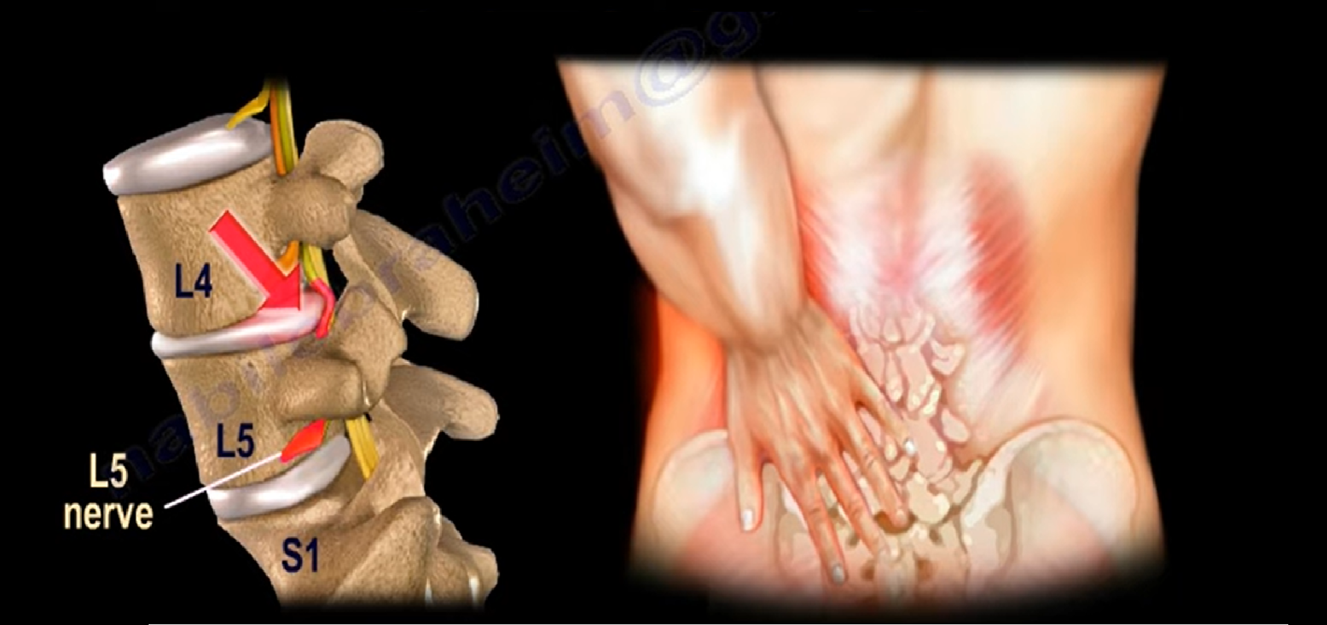
Profile of the “Ideal” Low Back Pain Patient
-
Clinical Concept: Most low back pain resolves with conservative management and time.
-
The “Ideal” Low Back Pain Patient Profile:
-
Acute onset of non-traumatic axial low back pain.
-
Pain can be severe and incapacitating.
-
May have limited lumbar range of motion.
-
Absence of distal radiation: No pain radiating below the knee level.
-
Absence of red flags: No constitutional symptoms (fever, chills, night sweats, unexplained weight loss).
-
Absence of neurological compromise: No bowel/bladder dysfunction, sensory loss, weakness, or pathologic upper motor neuron reflexes.
-
Pain improves with short periods of rest.
-
-
Axial vs. Radicular Pain Dominance:
-
Patients with back pain greater than leg pain have an excellent prognosis with conservative care.
-
Patients with leg pain greater than back pain have true sciatica/radiculopathy, requiring a more cautious diagnostic approach.
-
Natural History & Recovery Timeline
-
1 Week: Pain resolves in approximately 50% of patients with simple low back pain.
-
4 Weeks (1 Month): Acute axial pain resolves in 90% of patients.
-
6 Weeks: Nearly all uncomplicated, non-specific low back pain episodes subside.
-
Treatment Principles:
-
Avoid bed rest.
-
Prescribe short-term symptomatic relief (limited analgesics, NSAIDs, short-course muscle relaxants).
-
Reassure the patient regarding the favorable natural history.
-
Encourage early mobilization and return to normal daily activities as tolerated.
-
The most effective treatment for non-specific low back pain is an early return to work.
-
Occupational & Lifestyle Risk Factors
-
Poor physical conditioning and sedentary lifestyle.
-
Cigarette smoking.
-
Occupational tasks involving repetitive forward bending, stooping, or heavy lifting.
-
Occupational exposure to prolonged whole-body vibration (e.g., heavy machinery or professional driving).
Imaging Recommendations and Asymptomatic Imaging Pitfalls
-
Timing of Routine Imaging: Diagnostic imaging (plain radiographs, CT, or MRI) is not indicated within the first 6 weeks for uncomplicated, non-traumatic low back pain.
-
The Radiograph Paradox: Routine X-rays early in an uncomplicated course may improve short-term patient satisfaction, but they do not improve clinical outcomes.
-
Indications for Early Imaging:
-
Red flags: suspicion of trauma, occult fracture, neoplasm/metastasis, or infectious spondylodiscitis.
-
Progressive or severe neurological deficits.
-
Persistent, unremitting symptoms failing conservative management beyond 6 weeks.
-
-
Structural Findings on Plain X-rays:
-
Spondylolisthesis or segmental instability.
-
Lumbar spinal stenosis.
-
Degenerative spondyloarthropathy and severe structural deformities.
-
High False-Positive Rate of Lumbar Spine MRIs
-
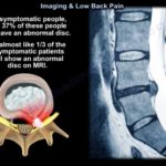
Asymptomatic Age Group Under 40 Years: Approximately 30% of healthy, pain-free subjects exhibit intervertebral disc abnormalities or protrusions on cross-sectional MRI.
-
Asymptomatic Age Group Over 60 Years: Roughly 90% of pain-free subjects demonstrate degenerative disc changes or canal narrowing on MRI.
-
Key Takeaway: An isolated MRI finding of a disc bulge or protrusion is not an automatic indication for surgery. Always correlate imaging with clinical and neurological findings.
Diagnostic Accuracy: Sciatica and Disc Herniations
-
Pathophysiology of Disc-Related Pain:
-
True sciatica represents mechanical compression and chemical irritation of a lumbosacral nerve root.
-
Pain worsens with maneuvers that increase intradiscal pressure or stretch neural structures: sitting, coughing, sneezing, and forward trunk flexion.
-
-
Straight Leg Raise (SLR) Test:
-
Performed by passively raising the patient’s leg with the knee fully extended.
-
Presence of radicular pain reproduced between 30° and 70° indicates dynamic nerve root tension.
-
An isolated positive SLR correlates with an underlying disc herniation in approximately 66% of patients.
-
Diagnostic Hierarchy for Lumbar Disc Herniation
-
Isolated Physical Finding (Positive SLR alone): 66% diagnostic accuracy.
-
Physical Finding + Confirmatory MRI (Positive SLR + Disc on MRI): Diagnostic accuracy increases to 86%.
-
Complete Clinical Triad (Neurological Deficit + Positive SLR + Disc on MRI):
-
Includes objective motor weakness, dermatomal sensory loss, or diminished deep tendon reflexes.
-
Achieves a 95% diagnostic accuracy for disc herniation.
-
Predicts a 95% surgical success rate following lumbar decompression/discectomy.
-
High-Yield Clinical Summary
-
Acute axial low back pain without distal radiation or red flags is self-limiting and does not require early imaging.
-
Early return to work and daily activity provides better recovery than bed rest.
-
High rates of disc abnormalities on MRI in asymptomatic individuals mean imaging must always be interpreted alongside clinical findings.
-
The combination of an objective neurological deficit, a positive Straight Leg Raise test, and a concordant MRI achieves the highest diagnostic accuracy (95%) and surgical predictability for disc herniation.


Leave a Reply