Courtesy: Prof Nabil Ebraheim, University of Toledo, Ohio, USA
Basic Facts
- Commonly seen in children aged 10–15 years
- Rare below 3 years
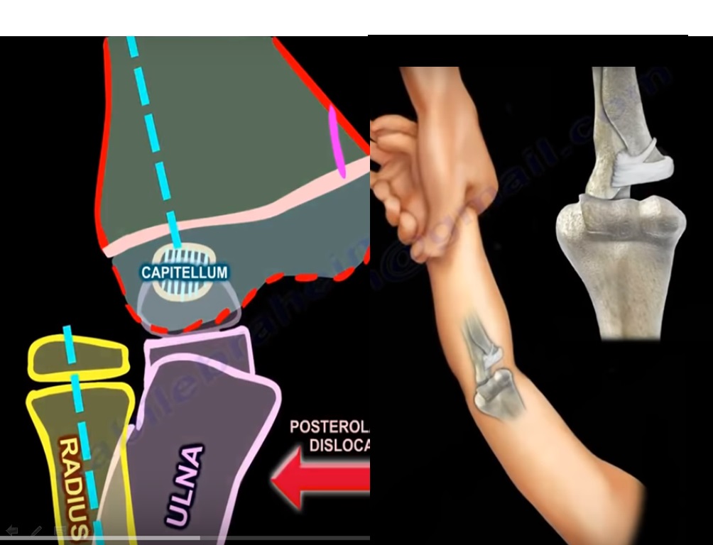
- Most common type: posterolateral dislocation
- Relationship between radius and ulna is maintained
- This helps distinguish it from Monteggia fracture-dislocation
Treatment of Elbow Dislocation
- Closed reduction
- Early mobilization
Always Assess For
- Medial epicondyle fracture
- Intra-articular entrapment of fracture fragment
Indications for Surgery (ORIF)
- Fragment entrapped within the joint
- Significant displacement
Conditions Associated With or Confused With Elbow Dislocation
1. Medial Epicondyle Fracture
- Commonly associated with elbow dislocation
- Fragment may be trapped within the joint, leading to incongruity
- Can be difficult to identify, especially after spontaneous reduction
Management
- ORIF when:
- Fragment is entrapped
- Significant displacement is present
2. Nursemaid’s elbow
- Age group: 2–3 years
- Cause: traction injury
Pathology
- Annular ligament slips, resulting in subluxation of the radial head
Clinical Features
- Child refuses to use the arm
- Arm held in slight flexion and pronation
- X-ray appears normal
Treatment
- Reduction using supination and flexion
- Immobilization is not required
3. Congenital Radial Head Dislocation
- Usually bilateral
- No history of trauma
Findings
- Posterior dislocation
- Hypoplastic capitellum
- Radius appears bowed and shortened
Key Point
- Not reducible
Treatment
- Radial head excision in adulthood if symptomatic
4. Monteggia Fracture-Dislocation
- Ulna fracture associated with radial head dislocation
Key Diagnostic Feature
- Radiocapitellar line is disrupted
Clinical Points
- Frequently missed injury
- Most common type involves anterior radial head dislocation
Complication
- Posterior interosseous nerve injury
Treatment
- Anatomical reduction of the ulna
If Missed
- Ulnar osteotomy with open reduction
5. Transphyseal Separation of Distal Humerus
- Seen in very young children, typically below 2–3 years
- Often mistaken for elbow dislocation
Key Differences
| Feature | Elbow Dislocation | Transphyseal Separation |
|---|---|---|
| Age | Older children | Infants |
| Displacement | Posterolateral | Posteromedial |
| Radiocapitellar line | Disrupted | Maintained |
| Capitellum | Visible | Not ossified |
Important Consideration
- Always evaluate for possibility of non-accidental trauma
Key Radiological Rule
- Radiocapitellar line:
- Should pass through the capitellum in all views
Clinical Use
- Helps differentiate:
- Elbow dislocation
- Monteggia lesion
- Transphyseal injury
Exam Pearls
- Elbow dislocation is rare below 3 years, consider transphyseal separation
- Entrapped medial epicondyle requires surgical treatment
- Normal X-ray with refusal to use arm suggests nursemaid’s elbow
- Always assess radiocapitellar alignment
- Missed Monteggia lesions can lead to major complications

Leave a Reply