
Courtesy Dr. Abhay Nene, Dr Ashok Shyam, Ortho TV
Osteoporotic Vertebral Compression Fractures: Vertebroplasty vs. Medical Management
1. Systemic Pathology & Core Principles
-
Osteoporosis as a Systemic Disease:
-
Osteoporosis is a generalized systemic disease; management must target the patient holistically rather than isolating the index fracture.
-
Orthopaedic oversight often mirrors fixing an intertrochanteric fracture while ignoring osteoporosis, analogous to treating spinal tuberculosis without anti-tubercular therapy (ATT).
-
-
Biomechanics of Vertebroplasty:
-
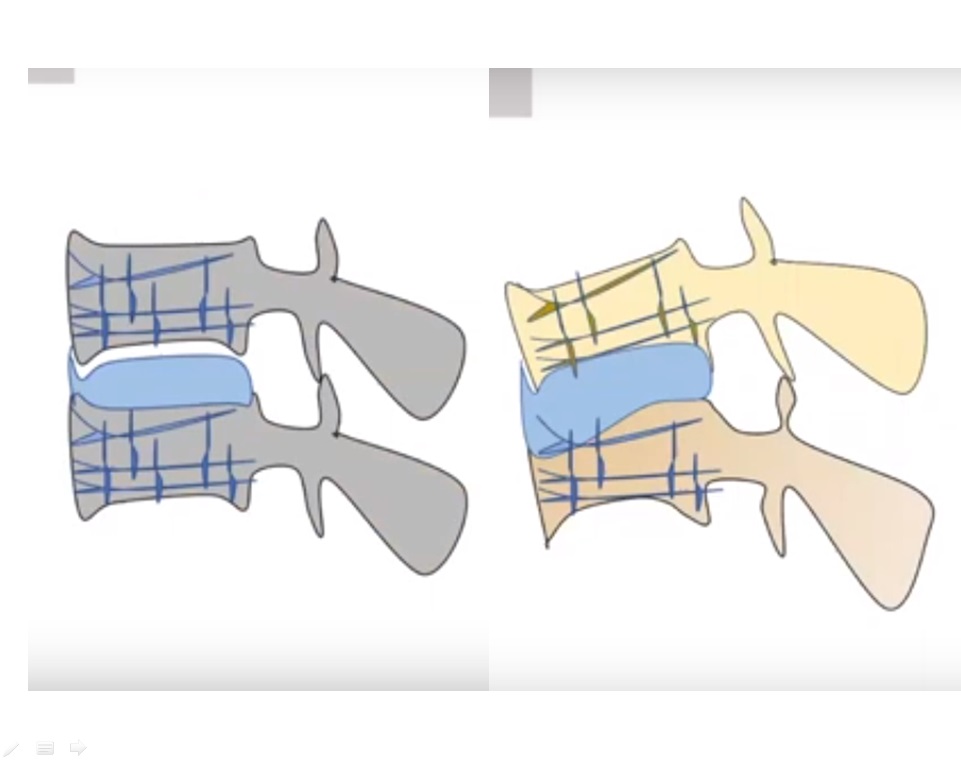
Introduces a rigid polymethylmethacrylate (PMMA) block into structurally weak, porous bone (“a stone in a pile of cushions”).
-
Provides immediate local structural stability and pain relief, but creates a severe stress riser due to mismatched Young’s modulus of elasticity.
-
Longer-term follow-up beyond 3 to 4 months frequently demonstrates structural breakdown, progressive collapse, and eggshell crackling around the cement.
-
2. Imaging Protocol & Contraindications to Vertebroplasty
Plain radiographs alone are insufficient for surgical planning; dedicated MRI and CT evaluation are mandatory.
-
Neural Canal Encroachment:
-
Plain radiographs cannot reliably confirm spinal canal compromise.
-
MRI is necessary to evaluate posterior superior cortical fragments; pressurising PMMA risks pushing retro-pulsed fragments further into the neural canal, creating new neurological deficits.
-
-
Non-Contained Fractures & Endplate Incompetence:
-
CT scans detect cortical defects and leakage paths near the endplates.
-
Breach of the endplate leads to intradiscal cement extrusion, inducing persistent disc-vertebral instability (a soft-tissue instability that cannot unite conservatively and demands surgical reconstruction).
-
-
Significant Kyphotic Deformity:
-
Pathological sagittal alignment concentrates high biomechanical loads across the anterior column; cement augmentation in this setting accelerates construct collapse.
-
-
Posterior Column / Ligamentous Instability:
-
Fractures appearing as simple compression injuries may conceal posterior element disruption.
-
Indicated clinically by localized posterior midline tenderness and radiologically by hyperintensity/disruption of the posterior ligamentous complex (PLC) on MRI (“white line”), necessitating open surgical stabilization rather than cement augmentation.
-
-
Pre-existing Adjacent Pathology & Severe Osteoporosis:
-
Highly prevalent in multiple multilevel osteoporotic compression fractures.
-
-
High Physical Demand in Elderly Patients:
-
Active lifestyle demands subject the cemented level to repetitive cyclical loading, causing the rigid PMMA block to erode/dent adjacent trabecular bone and precipitating secondary failure.
-
3. Cellular & Biomechanical Basis of Pharmacotherapy
-
Pathophysiological Basis of Osteoporosis:
-
Defined by trabecular structural architectural loss/defective trabeculae, not merely deficient mineral content (which characterizes osteomalacia).
-
-
Limitations of Bisphosphonates:
-
Act primarily by hyper-mineralizing residual bone matrix without regenerating lost microarchitecture.
-
Increases bone mineral density (BMD) on DEXA scans, but yields brittle, hyper-dense bone with compromised elasticity, contributing to atypical subtrochanteric fractures.
-
-
Anabolic Actions of Teriparatide:
-
Functionally stimulates de novo trabecular osteogenesis to restore structural connectivity and microarchitecture.
-
Reconstructs bone stock without creating focal mechanical stress risers, outperforming purely mechanical/surgical augmentation strategies.
-
4. Clinical Rescue Scenarios for Teriparatide
-
Vertebral Compression Fractures with Incipient Neurology:
-
Successfully bails out non-operative patients or revision scenarios (e.g., adjacent fractures following transforaminal lumbar interbody fusion [TLIF]) unwilling to undergo repeat surgery.
-
-
Failed Vertebroplasty:
-
Reverses peri-cement osteolysis and mechanical erosion of adjacent host bone where salvage surgery offers poor purchase.
-
-
Complicated / Disaster Revision Spine Surgery:
-
Rescues structural failures and hardware complications secondary to poor osteoporotic bone quality.
-
Master Clinical Correlates
-
Systemic vs. Local Focus: Always prioritize medical osteoporosis management over isolated local stabilization.
-
Imaging Algorithm: MRI assesses neural canal status and PLC integrity; CT rules out non-contained endplate/posterior wall defects.
-
Biomechanical Pitfall: Vertebroplasty alters the modulus of elasticity, creating stress risers and adjacent collapse.
-
Bisphosphonates: Increase mineral density without restoring trabecular continuity, leading to brittle bone matrix.
-
Teriparatide: Induces true trabecular osteogenesis; indicated as primary treatment for severe osteoporosis and as a rescue agent for failed vertebroplasty or hardware breakdown.
-
Clinical Recommendation: Routine medical optimization with teriparatide for eligible osteoporotic patients; reserve vertebroplasty strictly for highly selected indications.

Leave a Reply