
Courtesy: Prof Nabil Ebraheim, University of Toledo, Ohio, USA
Transient Osteoporosis of the Hip vs Osteonecrosis of the Femoral Head
1. Transient Osteoporosis of the Hip
Transient osteoporosis is not osteonecrosis of the femoral head.
Typical Patients
It usually affects:
- Pregnant women
- Men in the fifth decade of life
Clinical and Radiographic Features
A characteristic feature is that the symptoms are more significant than the X-ray findings.
Plain radiographs may show:
- Osteopenia
MRI is much more characteristic.
MRI Findings
MRI shows:
- Bone marrow edema
- Increased T2 signal intensity
The signal abnormality may involve:
- Femoral head
- Femoral neck
- Sometimes the intertrochanteric region
An important distinction is:
Transient osteoporosis does not show the double-line sign characteristic of osteonecrosis.
Treatment
Transient osteoporosis:
- Is not a tumor
- Is not osteonecrosis
- Does not require surgery
2. Osteonecrosis of the Femoral Head
Osteonecrosis is a fundamentally different condition.
Bilateral Disease
Osteonecrosis may be bilateral in approximately 80% of patients according to the lecture.
Therefore:
Always assess the opposite hip, even if it is asymptomatic.
3. Importance of Early Diagnosis
Early diagnosis may improve the possibility of successful head-preserving treatment.
Examples mentioned include:
- Core decompression
- Bone grafting
In advanced disease:
Femoral head collapse
can occur.
Once significant collapse has occurred, head-preserving treatment is unlikely to provide a good outcome, and the femoral head may need to be replaced.
4. Imaging of Osteonecrosis
Plain Radiographs
Obtain:
- AP view
- Frog-leg lateral view
The frog-leg lateral view may demonstrate the characteristic crescent sign associated with subchondral fracture.
However, early osteonecrosis may have normal radiographs.
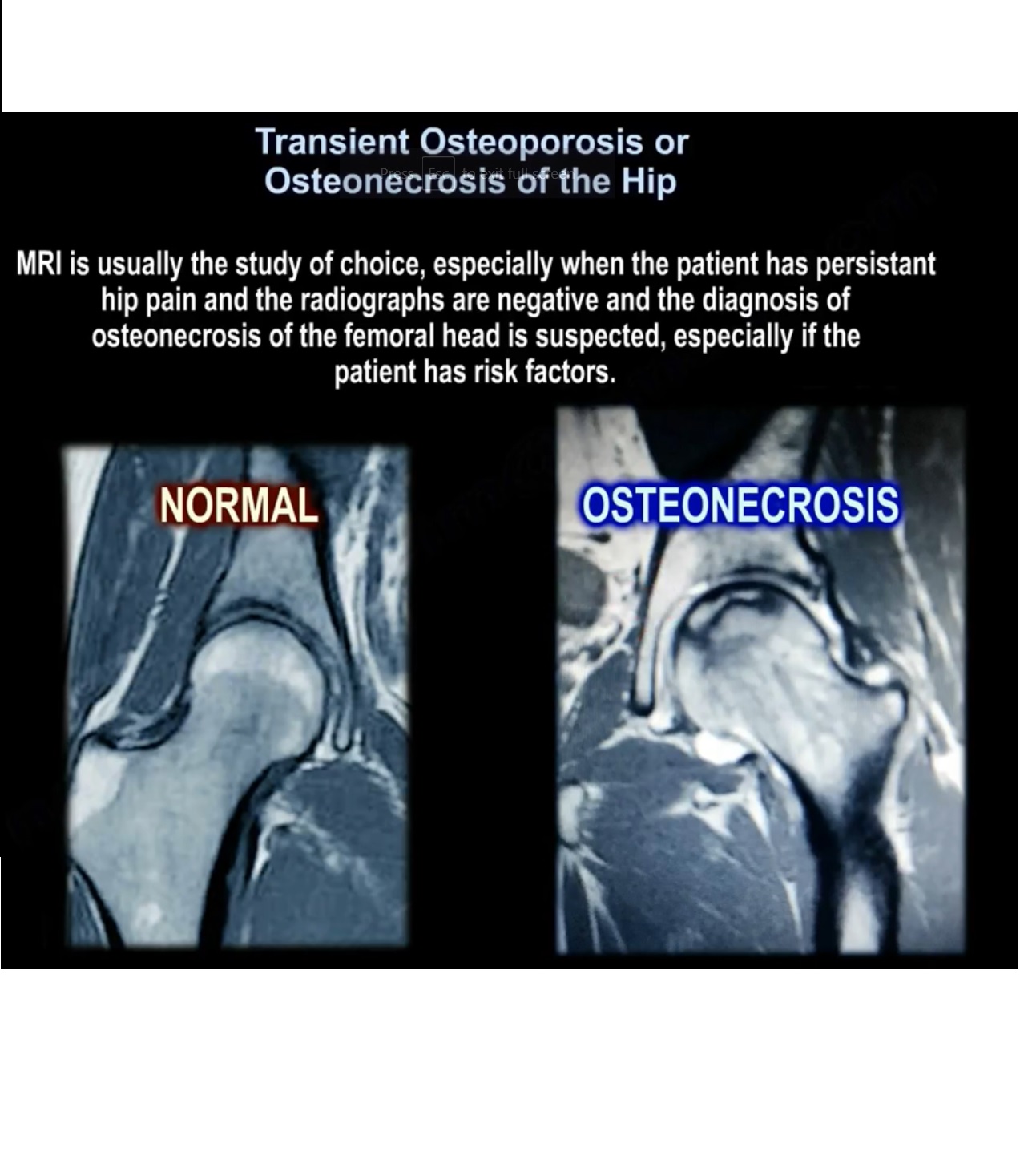
5. MRI
MRI is the study of choice, particularly when:
- Hip pain persists
- Radiographs are negative
- Osteonecrosis is suspected
- Risk factors are present
T1 MRI
A typical finding is:
- Well-defined band of low signal intensity
- Usually in the superior/anterior portion of the femoral head
This represents the interface between necrotic and reparative bone.
6. The Double-Line Sign
The double-line sign is best appreciated on T2-weighted MRI.
It consists of:
- An inner high-signal line
- An outer low-signal line
The high-signal component represents hypervascular granulation tissue.
The low-signal peripheral line represents the reactive interface.
This creates the characteristic:
Double-line sign
The double-line sign is an important MRI feature distinguishing osteonecrosis from transient osteoporosis.
7. Size of the Lesion Matters
The size of the osteonecrotic lesion is an important factor in:
- Development of symptoms
- Disease progression
- Prognosis
Better Prognosis
The best prognosis occurs with:
- Small lesion
- Sclerotic margins
Bone Marrow Edema
The presence of bone marrow edema on MRI is associated with:
- Worsening pain
- Future disease progression
8. Multifocal Osteonecrosis
Multifocal osteonecrosis refers to disease involving three or more skeletal sites.
Possible sites include:
- Hip
- Knee
- Shoulder
- Ankle
The lecture states that multifocal disease occurs in approximately 3% of patients.
Important Clinical Point
If a patient presents with osteonecrosis at a site other than the hip:
MRI of the hip should be considered to look for an asymptomatic femoral-head lesion.
Quick Comparison
| Feature | Transient Osteoporosis | Osteonecrosis |
|---|---|---|
| Basic pathology | Transient osteoporosis | Bone necrosis |
| Typical patients | Pregnancy, men in 5th decade | Various risk groups |
| X-ray | Often normal or osteopenic | May be normal early |
| MRI | Diffuse bone marrow edema | Characteristic low-signal band |
| Femoral head/neck edema | Common | May occur |
| Double-line sign | Absent | Present |
| Crescent sign | Not characteristic | May indicate subchondral fracture |
| Bilateral disease | Not emphasized | Common |
| Early treatment | Non-operative | Head-preserving procedures may be possible |
| Late collapse | Not expected as osteonecrosis | Femoral-head collapse |
| Advanced treatment | Usually no surgery | Femoral-head replacement may be required |
Exam Pearls
- Transient osteoporosis is not osteonecrosis.
- Transient osteoporosis often has more symptoms than X-ray abnormalities.
- MRI in transient osteoporosis shows bone marrow edema without a double-line sign.
- Osteonecrosis may be bilateral, so examine the opposite hip.
- MRI is the investigation of choice when radiographs are negative but osteonecrosis is suspected.
- Crescent sign = subchondral fracture.
- Double-line sign on T2 MRI = osteonecrosis.
- Lesion size is an important prognostic factor.
- Bone marrow edema is associated with worsening pain and progression.
- Early osteonecrosis may be amenable to core decompression or bone grafting; advanced collapse may require femoral-head replacement.


Leave a Reply