• Skip to main content
  • Skip to secondary menu
  • Skip to primary sidebar
OrthopaedicPrinciples.com

OrthopaedicPrinciples.com

Integrating Principles and Evidence

Integrating Principles and Evidence

  • Home
  • Editorial Board
  • Our Books
    • Evidence Based Orthopaedic Principles
  • Courses
  • Exams
  • Reviews
  • Live Program
  • Contact

Osteoid Osteoma

Courtesy: Dr Amr Abdelgawad,
Texas Tech University, El Paso, USA

This video describes about the benign bone tumor osteoid osteoma.

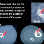
  • It is characterized by presence of a nidus (bone forming area) surrounded by reactive area of thickened bone . It is more common in 2nd & 3rd decades, usually affecting men.
  • Common site is the diaphysis of long bones. when it affects spine, it can result in scoliosis
  • Patient usually presents with pain which is more during night ,relieved by aspirin & other NSAID’s & not relieved by rest.
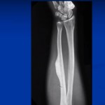
  • X-ray shows a radiolucent area surrounded by reactive dense sclerotic bone
  • CT scan is better than MRI in diagnosing osteoid osteoma as the nidus can’t be seen accurately due to soft tissue oedema.
  • CT shows radiolucent area surrounded by dense hypersclerotic bone. Here the nidus is less than 1.5 cm. If it is more than 1.5 cm it will fall into the category of osteoblastoma
  • Osteoid osteoma can be seen as region of hotspot in bone scan.
  • Medical treatment is by the use of NSAIDs
  • Surgical treatment includes excision of the lesion with nidus & CT guided percutaneous radio frequency ablation

Osteoid osteoma

Post Views: 3,521

Related Posts

  • Osteoid Osteoma: Clinical Features and Management

    Courtesy: Prof Nabil Ebraheim, University of Toledo, Ohio, USA

  • Benign Bone Forming Tumours

    Courtesy: Dr James Wittig, Orthopaedic Oncologist, Sarcoma Surgeon wwww.tumorsurgery.org Books by Dr James Wittig:-

  • Musculoskeletal Oncology Fellowship

    Musculoskeletal Tumours Fellowship Aims:The fellow will be involved in the management of patients presenting to…

Reader Interactions

Leave a Reply

Your email address will not be published. Required fields are marked *

Primary Sidebar

Follow Us

instagram slideshare

Categories

  • -Applied Anatomy
  • -Approaches
  • -Basic Sciences
  • -Cartilage & Meniscus
  • -Classifications
  • -Examination
  • -Foot and Ankle
  • -Foot and Ankle Trauma
  • -FRCS(Tr and Orth) tutorials
  • -Gait
  • -Hand and Wrist
  • -Hand and Wrist Trauma
  • -Hand Infections
  • -Hip and Knee
  • -Hip Preservation
  • -Infections
  • -Joint Reconstruction
  • -Knee Arthroplasty
  • -Knee Preservation
  • -Metabolic Disorders
  • -Oncology
  • -OrthoBiologics
  • -OrthoPlastic
  • -Paediatric Orthopaedics
  • -Paediatric Trauma
  • -Patellofemoral Joint
  • -Pelvis
  • -Peripheral Nerves
  • -Principles
  • -Principles of Surgery
  • -Radiology
  • -Rheumatology
  • -Shoulder and Elbow
  • -Shoulder and Elbow Arthroplasty
  • -Spine Deformity
  • -Spine Oncology
  • -Spine Trauma
  • -Spine, Pelvis & Neurology
  • -Sports Ankle and Foot
  • -Sports Elbow
  • -Sports Knee
  • -Sports Medicine
  • -Sports Medicine Hip
  • -Sports Shoulder
  • -Sports Wrist
  • -Statistics
  • -Technical Tip
  • -Technology in Orth
  • -Trauma
  • -Trauma (Upper Limb)
  • -Trauma Life Support
  • -Trauma Reconstruction
  • Book Shelf
  • Book Shelf Medical
  • Careers
  • Case Studies and Free Papers
  • DNB Ortho
  • Evidence Based Orthopaedic Principles
  • Evidence Based Orthopaedics
  • Exam Corner
  • Fellowships
  • Guest Editor
  • Guest Reviews
  • Image Quiz
  • Instructional Course Lectures
  • Journal Club
  • MCQs
  • Meetings and Courses
  • Multimedia
  • News and Blog
  • Plaster Techniques
  • Podcasts
  • Public Health
  • Rehabilitation
  • Research
  • Shorts and Reels
Copyright@orthopaedicprinciples.com. All right rerserved.