Courtesy; Prof Nabil Ebraheim, University of Toledo, Ohio, USA
1. Common Pediatric Elbow Fractures
A. Supracondylar fracture of humerus
- Most common pediatric elbow fracture
- Most common cause of elbow effusion in children
Diagnosis
- Anterior humeral line assessment
B. Fractures Involving Ossification Centers
Elbow ossification centers CRITOE
| Structure | Age (years) |
|---|---|
| Capitellum | 1 |
| Radial head | 3 |
| Medial epicondyle | 5 |
| Trochlea | 7 |
| Olecranon | 9 |
| Lateral epicondyle | 11 |
Key Point
- Essential for interpreting pediatric elbow X-rays
1. Transepiphyseal Separation – Distal Humerus
Key Features
- Age below 1 year
- Always consider non-accidental injury
- Often mistaken for elbow dislocation
Differentiation from Dislocation
- Radiocapitellar alignment maintained
- Olecranon displacement pattern:
- Dislocation: posterior and lateral
- Transepiphyseal separation: posterior and medial
2. Lateral condyle fracture of humerus
Characteristics
- Usually Salter-Harris Type IV
- Most important intra-articular fracture in children
Management
- Internal oblique view required even if undisplaced
- Close follow-up due to risk of displacement
- Majority require surgical fixation using lateral approach
Avoid
- Posterior approach due to risk of avascular necrosis
Complications
- Non-union leading to cubitus valgus
- Late ulnar nerve palsy
Management of Complications
- Bone grafting for non-union
- Ulnar nerve transposition if symptomatic
3. Medial epicondyle fracture
Key Points
- Last ossification center to fuse
- Commonly associated with elbow dislocation
Clinical Concern
- Fragment may be incarcerated within the joint
Management
- Usually conservative
Indications for Surgery
- Displacement greater than 1 cm
- Fragment trapped within the joint
4. Radial head fracture / Neck Fracture
Management Based on Angulation
- Less than 30 degrees: conservative
- Around 30 degrees: closed reduction
- Residual angulation greater than 45 degrees: open reduction
Techniques
- Percutaneous pin used as joystick
Complications
- Radioulnar synostosis
- Osteonecrosis
- Loss of motion
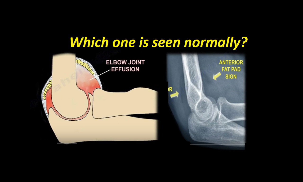
2. Occult Elbow Injury (Fat Pad Sign)
Concept
- Joint effusion displaces fat pads, making them visible on X-ray
Fat Pad Findings
Anterior Fat Pad
- Normal: small and parallel to humerus
- Abnormal: elevated triangular appearance (sail sign)
Posterior Fat Pad
- Normally not visible
- If visible, always pathological
Key Rule
- Visible posterior fat pad indicates occult fracture until proven otherwise
Common Causes
Children
- Most commonly supracondylar fracture
Adults
- Most commonly radial head or neck fracture
Why Fracture May Be Missed
- Undisplaced fracture
- Extra-articular fracture component
- Poor radiographic quality
Special Imaging View
Radiocapitellar (Greenspan) View
- Elbow flexed to 90 degrees
- X-ray beam angled 45 degrees proximally
- Useful for detecting radial head and neck fractures
3. Systematic Approach to Pediatric Elbow X-ray
Four Key Steps
- Ensure good quality X-ray
- Correlate with age and CRITOE sequence
- Look for fractures or missing ossification centers
- Assess fat pad signs
4. Important Radiological Lines
A. Anterior Humeral Line
- Should pass through middle third of capitellum
Clinical Use
- Diagnosis of supracondylar fractures
- Helps identify mechanism (flexion or extension type)
B. Radiocapitellar Line
- Line drawn along radial neck
- Should intersect capitellum in all views
Clinical Use
- Detect radial head dislocation
- Differentiate transepiphyseal separation
Exam Pearls
- Posterior fat pad indicates occult fracture
- Most common fracture is supracondylar fracture
- Most important intra-articular fracture is lateral condyle fracture
- CRITOE sequence is essential for interpretation
- Medial epicondyle fracture with dislocation may have trapped fragment
- Open reduction of radial neck fractures has high complication rates
Final Takeaway
- Pediatric elbow evaluation depends on:
- Age
- Ossification pattern
- Radiographic alignment
- Missing subtle signs leads to:
- Malunion
- Deformity
- Long-term functional impairment

Leave a Reply