Courtesy: John Ebnezar, Bangalore
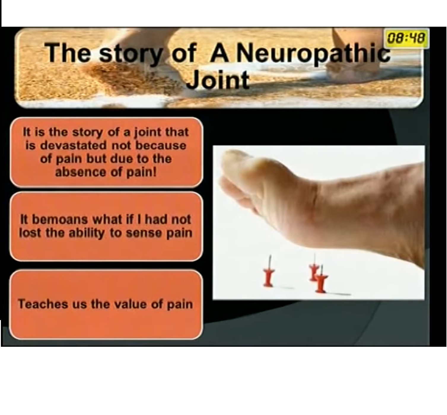
NEUROPATHIC JOINT
- A neuropathic joint also known as Charcot’s joint is a progressive and degenerative condition characterised by joint destruction due to loss of normal of sensory innervation.
- This loss of sensation leads to repetitive trauma and microfractures resulting in joint deformity and instability .
Etiology:
Can results from various conditions-
1. Diabetes Mellitus
2. Syringomyelia
3. Tabes dorsalis
4. Leprosy
5. Spinal cord injuries
Joints involved-
1. TMT – 60%
2. MTP
3. Ankle joint
4. Knee joint – rare
5. Shoulder joint – Syringomyelia
6. Hip joint
Clinical presentation:
• Unilateral
• Painless loss of function
• Erythema
• Edema
• Increased temperature
• Unstable ,swollen joint
• Plantar ulcers
Distribution-
- Leading cause of Charcot’s foot- Diabetes Mellitus
- Mid arch is the most common area involved.
Differential Diagnosis-
- Osteomyelitis
- Osteoarthritis
Investigations :
Xray– findings
- Atrophic –osteolysis of distal metatarsals in forefoot
- “licked candy stick “ appearance seen at distal aspect of metatarsal.
- Hypertrophic – characterised by Acute periarticular fracture and joint dislocation.
Bone scans –
- Sensitive indicator of hyperemia
- Surface skin temperature
MRI- features
- Destruction , Dislocation,Distention- edema
Brodsky Classification –(based on location of Charcot’s joint)
• Type 1- Lisfrank joint 26-60%
• Type 2- Chopart’s joint and subtalar joint 30-35%
• Type 3A – Ankle joint
Type 3B- Posterior calcaneous
• Type 4- Multiple regions of foot and or ankle
• Type 5- Forefoot
Treatment
• Acute inflammatory stage –immobilisation
• Resolution stage –accommodative care
1.inlay depth shoes
2.accomodative depth orthoses
3.ankle foot orthoses
• Plantigrade surface not achieved-
Then surgical stabilisation or reconstruction
Non operative measures :
• Activity modification
• Avoid weight bearing
• Total contact casting / bracing
SURGERIES IN CHARCOT FOOT:
1.Exostectomies
2.Reconstruction or Reshaping
- Reshaping of foot eliminates a bony prominence on top or bottom of foot.
3. Limb salvage procedures
• Midfoot arthrodesis
• Triple arthrodesis
• Tibiocalcaneal arthrodesis
• Ankle arthrodesis – for non braceable neuropathic ankle deformity.
ankle involvement leads to ulceration ,osteomyelitis and amputation
Arthrodesis before the ulcerated lesion appears is considered as a limb salvage treatment.
4. External Fixation
- For selected patients ,external fixation after surgical debridement ,considered a reasonable alternarive to below knee amputation.
5. Below knee amputation
When to consider Charcot’s neuropathy?
• Diabetic patient
• Inflamed foot
• Absence of fever
• Elevated CRP or ESR
• In above condition, infection is highly unlikely – hence Charcot process must be considered primarily.
Lecture very informative, speaker, could really do with slowing down slightly , I found the instruction well planned . With good delivery .