Courtesy: Prof Nabil Ebraheim, University of Toledo, Ohio, USA
1. Tarsal Tunnel Syndrome
Overview
-
One of the most common nerve injuries in the lower extremity.
-
Approximately 80% of cases have no identifiable cause.
-
Involves compression of the posterior tibial nerve.
Symptoms
-
Burning sensation
-
Numbness
-
Tingling
-
Electric shock–like pain
-
Symptoms occur on the plantar (bottom) aspect of the foot
Persistent Symptoms After Surgery
-
If symptoms persist six months after surgery:
-
Likely cause is incomplete release of impinging structures.
-
-
Recurrence is usually due to:
-
Inadequate decompression.
-
-
Repeat surgery is generally not advisable.
-
Surgical decompression is not as predictably successful as carpal tunnel release.
-
Outcomes are better when the cause is a ganglion compared to other causes.
Anatomy Behind the Medial Malleolus (“Tom, Dick, and Harry”)
Structures passing behind the medial malleolus:
-
Tom – Tibialis posterior
-
Dick – Flexor digitorum longus
-
Posterior tibial artery
-
Posterior tibial vein
-
Nerve – Posterior tibial nerve
-
Harry – Flexor hallucis longus (important muscle of the foot)
2. L5 Nerve Root
-
Supplies extension of the big toe.
-
Extensor hallucis longus is supplied by the L5 nerve root.
-
Should not be confused with flexor hallucis longus.
3. Cutaneous Innervation of the Foot
Plantar Surface
-
Medial plantar nerve
-
Lateral plantar nerve
-
Sural nerve (lateral side)
-
Saphenous nerve (medial side)
Important distinctions:
-
Medial side of the foot ? Saphenous nerve
-
Lateral side ? Sural nerve
-
Medial plantar nerve and lateral plantar nerve supply the plantar surface.
Cutaneous innervation differs from the dorsum (top) of the foot.
4. Lumbar Disc Herniation and Sensory Changes
Posterolateral L5–S1 Disc Herniation
-
Decreases sensation over the lateral aspect of the foot.
-
Corresponds to S1 distribution.
Foraminal L5–S1 Disc Herniation
-
Affects the L5 nerve root.
-
Decreases sensation over the dorsum (top) of the foot.
Sensory Summary
-
Top of the foot ? L5
-
Lateral side of the foot ? S1
-
Medial side of the foot ? L4
-
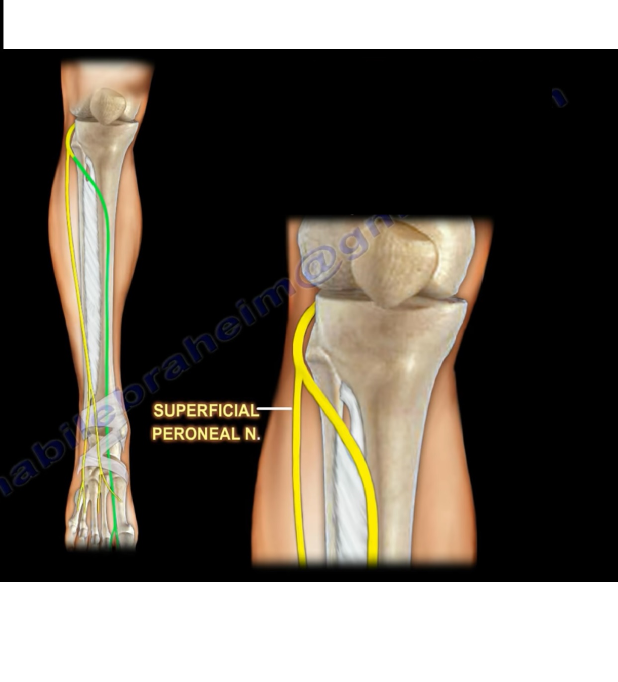
L5 sensory area on the dorsum corresponds to the superficial peroneal nerve distribution.
5. Sciatic Nerve Injury
Clinical Presentation
-
A high sciatic nerve lesion can mimic common peroneal nerve injury at the fibular head.
-
Both conditions may present with foot drop.
Differentiating Lesion Level
-
Short head of the biceps femoris:
-
Supplied by the common peroneal branch of the sciatic nerve.
-
-
Long head of the biceps femoris:
-
Supplied by the tibial branch of the sciatic nerve.
-
Role of EMG
-
EMG of the short head of the biceps femoris helps determine lesion location.
If EMG abnormal in short head:
-
Lesion is high (sciatic nerve level).
If EMG normal in short head:
-
Lesion is distal (common peroneal nerve at the fibular head).
This is an important examination point.
6. Deep Peroneal Nerve
-
Provides sensory supply to the first web space (between the first and second toes).
-
Superficial peroneal nerve supplies:
-
The remainder of the dorsum of the foot except the first web space.
-
Clinical relevance:
-
First web space sensation can be tested in anterior compartment syndrome, which most commonly affects the anterior compartment.
7. Lateral Plantar Nerve
-
Branch of the posterior tibial nerve.
-
Posterior tibial nerve arises from the tibial nerve.
-
Tibial nerve is a branch of the sciatic nerve.
Importance
-
Similar in function to the ulnar nerve in the hand.
-
Supplies most of the intrinsic muscles of the foot.
-
Supplies the interossei muscles.
-
Frequently tested in examinations.
Baxter’s Nerve
-
First branch of the lateral plantar nerve.
-
Can be confused with plantar fasciitis.
-
Involved in chronic heel pain.
-
Contributes to approximately 20% of heel pain cases.
-
Supplies the abductor digiti muscle.
-
May be injured during procedures involving insertion of a rod from the heel into the calcaneus and tibia.
8. Heel Pain
-
Multiple causes located within a small anatomical area.
-
Structures are close to each other, making diagnosis difficult.
-
Common causes include:
-
Plantar fasciitis
-
Baxter’s nerve involvement
-
9. Interdigital Neuroma (Morton’s Neuroma)
Definition
-
Compression of the interdigital nerve.
Location
-
Approximately 80% occur in the third interdigital space.
-
About 20% occur in the second interdigital space.
Symptoms
-
Localized plantar forefoot pain
-
Pain does not involve the entire foot.
-
Pain radiates distally in approximately 60% of cases.
-
Numbness occurs in approximately 40% of cases.
Examination Findings
-
Focal, localized tenderness in the plantar web space.
-
Tenderness is on the bottom (plantar side), not over the joint or dorsum.

Leave a Reply