Courtesy: Dr Strickland, Ashok Shyam TV, Ortho
Background and Evolution of Treatment
-
Medial patellofemoral ligament reconstruction was rarely performed two decades ago.
-
The ligament was historically under-recognized in patellar instability.
-
Over the last 12 to 13 years, understanding of its importance has significantly increased.
-
Patellar instability can lead to:
-
Loss of athletic participation for an entire season in approximately 20 percent of patients.
-
High incidence in young female athletes, including gymnastics, soccer, lacrosse, and football.
-
Significant disability with prolonged nonoperative management in many cases.
-
Associated Injuries and Long-Term Risks
-
Osteochondral or chondral injury is common following patellar dislocation.
-
Some patients progress to generalized knee osteoarthritis.
-
Instability symptoms do not necessarily correlate with severity of cartilage damage.
-
Careful evaluation is required in every case.
Historical Procedures
-
Several older surgical procedures for patellar instability demonstrated poor outcomes.
-
Some techniques have been abandoned due to high failure rates.
-
Modern treatment focuses on anatomical restoration and risk-based decision making.
Treatment Algorithm Overview
First-Time Dislocator
-
If cartilage injury is present:
-
Surgical management should be strongly considered.
-
Reconstruction of the medial patellofemoral ligament should be included.
-
-
If no cartilage injury:
-
Risk factors must guide decision-making.
-
High-risk patients should have a surgical discussion.
-
Multiple Dislocators
-
Surgical management is generally recommended.
-
Medial patellofemoral ligament reconstruction is indicated in most cases.
-
Nonoperative treatment is rarely appropriate after multiple instability episodes.
Predicting Recurrence
Certain patients have significantly higher recurrence rates.
High-Risk Features
-
Patella alta
-
Trochlear dysplasia
-
Age younger than 25 years
These patients may have:
-
Approximately 70 percent five-year recurrence risk.
Additional predictive factors:
-
Skeletal maturity
-
Trochlear dysplasia severity
-
Patella alta measurements
-
Contralateral patellar dislocation history
Patients with prior contralateral dislocation:
-
Three times higher risk of instability.
-
Prior instability in either knee increases recurrence risk significantly.
Special Considerations in Skeletally Immature Patients
-
Open growth plates limit surgical options.
-
Bony procedures are contraindicated.
-
Medial patellofemoral ligament reconstruction is the primary surgical option.
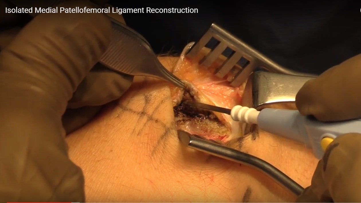
Isolated Medial Patellofemoral Ligament Reconstruction
-
In many cases, isolated reconstruction provides good short-term outcomes.
-
Published studies show favorable results at one to two years.
-
Long-term data beyond five years are critical for true success evaluation.
Clinical Examination Pearls
Patellar Tilt Assessment
-
Patient in full extension.
-
Both hands used to center patella.
-
Assess ability to evert lateral facet to neutral.
-
Tight lateral restraints may require lateral lengthening.
Important Warning
-
Do not perform lateral release or lengthening in patients with generalized ligamentous laxity (high Beighton score).
Femoral Tunnel Placement: Critical Factor
-
Most failures are due to incorrect femoral tunnel placement.
-
Incorrect positioning results in:
-
Non-isometric graft behavior.
-
Tunnel widening.
-
Overconstraint.
-
Poor clinical outcomes.
-
Key Principles
-
Start at the palpable sulcus.
-
Check graft isometry carefully.
-
Confirm positioning with fluoroscopy in every case.
-
Avoid placing the tunnel too high and too tight.
Improper placement may cause:
-
Excessive graft tension.
-
Patellar overload.
-
Catastrophic failure.
Patellar Fixation Pearls
-
Avoid violating patellar cortex.
-
Avoid damaging articular cartilage.
-
Incorrect anchor placement may lead to:
-
Patellar fracture.
-
Extensor mechanism compromise.
-
Severe patient dissatisfaction.
-
Preferred Technique
-
All-suture anchors placed carefully.
-
Avoid lateral facet violation.
-
Avoid excessive anterior cortex weakening.
Graft Fixation Technique Overview
-
Doubled graft attached to patella.
-
Femoral tunnel created after confirming isometry and fluoroscopic position.
-
Whip stitching of graft.
-
Button or adjustable fixation device allows tension fine-tuning.
-
Graft should not be overtightened.
-
Goal is appropriate stability, not excessive constraint.
Case Example: Skeletally Immature Patient
-
Young female patient with:
-
Patella alta
-
Trochlear dysplasia
-
High tibial tubercle to trochlear groove distance
-
-
Osteotomy desired but not possible due to open growth plate.
-
Large osteochondral fragment identified.
-
Surgical plan:
-
Medial patellofemoral ligament reconstruction.
-
Cartilage repair if fragment viable.
-
Fixation with absorbable implants when possible.
-
Cartilage Management Principles
-
If fragment viable:
-
Repair is preferred.
-
-
If fragment nonviable:
-
Consider cartilage restoration procedures.
-
-
Address cartilage injury at the index procedure whenever possible.
Key Surgical Pearls Summary
-
Open growth plate:
-
Only soft tissue procedures.
-
-
Isolated medial patellofemoral ligament reconstruction often effective.
-
Assess patellar tilt carefully.
-
Avoid lateral release in hyperlax patients.
-
Femoral tunnel accuracy is critical.
-
Avoid patellar cortex and cartilage violation.
-
Fine-tune graft tension.
Clinical Decision-Making Summary
-
Risk stratification improves recurrence prediction.
-
High-risk first-time dislocators should be counseled about surgery.
-
Multiple dislocators generally require reconstruction.
-
Cartilage injury mandates surgical consideration.
-
Early cartilage fixation improves long-term joint preservation.
Conclusion
-
Management of patellar instability has evolved significantly.
-
Medial patellofemoral ligament reconstruction is central to treatment.
-
Accurate surgical technique determines outcome.
-
Risk factor assessment allows individualized care.
-
Early and appropriate intervention may prevent recurrent instability and long-term degeneration.
Leave a Reply