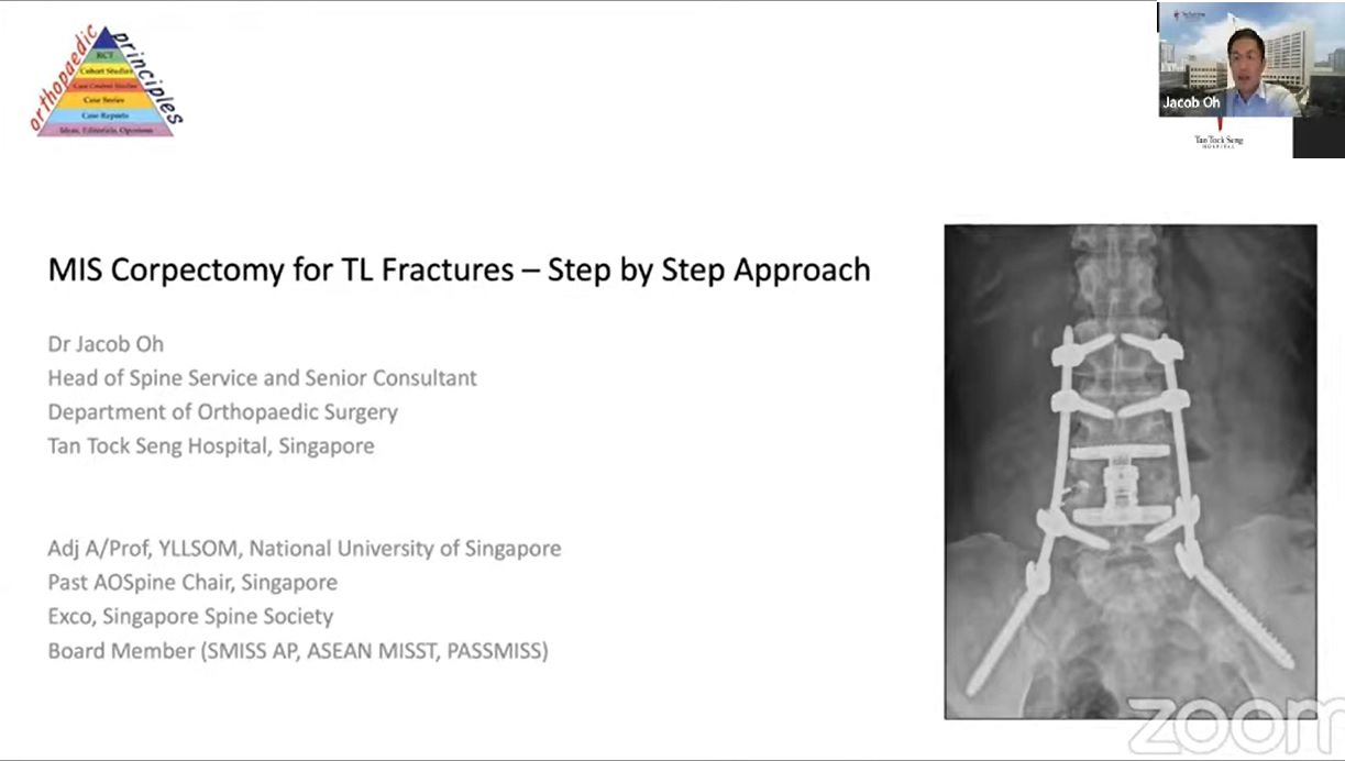
Courtesy: Jacob Oh, Chief of Spine Surgery, Tan Tock Seng Hospital, Singapore
Introduction
Osteoporotic vertebral compression fractures are becoming increasingly common due to the growing prevalence of osteoporosis. While many fractures can be managed conservatively or with posterior fixation alone, certain fracture patterns require anterior column reconstruction to achieve adequate stability, decompression, and long term outcomes.
This lecture discusses minimally invasive anterior corpectomy techniques for thoracolumbar fractures and demonstrates how these approaches can provide the advantages of anterior reconstruction while minimizing the morbidity traditionally associated with open anterior surgery.
Why Posterior Surgery Remains the Workhorse
For most thoracolumbar fractures, a posterior approach remains the preferred option because it is:
- Familiar and reproducible
- Technically straightforward
- Relatively fast
- Effective for decompression
- Suitable for emergency situations
Pedicle screw fixation remains the foundation of treatment for many spinal fractures.
When Anterior Column Support Becomes Necessary
Certain fractures cannot be adequately managed through a posterior approach alone.
Typical indications include:
- Vertebral body height loss greater than 50%
- Kyphotic angulation greater than 30°
- Severe anterior column destruction
- Large retropulsed fragments causing major canal compromise
- Osteoporotic Fracture (OF) Type 4 and Type 5 injuries
- Pathological vertebral body collapse
In these situations, anterior reconstruction provides:
- Superior anterior column support
- Improved fusion rates
- Direct neural decompression
- Ability to shorten posterior constructs
Anterior Approaches to the Spine
L5 Vertebral Body
Direct anterior approach
Similar to an anterior lumbar interbody fusion approach.
Key considerations:
- Mobilization of the iliac vessels and inferior vena cava
- Lateral access is limited by the iliac crest
L1 to L5 Vertebral Bodies
Lateral retroperitoneal approach
Commonly associated with XLIF and OLIF techniques.
Approach involves:
- Splitting abdominal wall musculature
- Entering the retroperitoneal space
- Accessing the vertebral body through an oblique corridor anterior to the psoas muscle
T11 and T12 Vertebral Bodies
Retropleural approach
The diaphragm becomes an important anatomical landmark.
- Structures below the diaphragm are approached retroperitoneally
- Structures above the diaphragm require retropleural or transthoracic access
Mid Thoracic Spine
Typically approached through:
- Right retropleural approach
- Right transthoracic approach
A right sided approach avoids the descending thoracic aorta.
Challenges of Traditional Anterior Surgery
Historically, anterior surgery has been associated with substantial morbidity.
Potential disadvantages
- Significant blood loss
- Difficult dural repair
- Major vascular injury risk
- Exposure related complications
- Limited multilevel access
- Need for assistance from vascular or thoracic surgeons
These concerns motivated the development of minimally invasive techniques.
Case 1: L4 Pathological Fracture from Metastatic Disease
Clinical Presentation
- 65 year old woman
- Breast cancer metastasis
- Severe back pain
- Neurogenic claudication
- Foot drop
- L4 pathological fracture with severe cauda equina compression
Surgical Strategy
- Retroperitoneal approach
- L4 corpectomy
- Direct decompression
- Cage reconstruction
- Posterior fixation
Step 1: Retroperitoneal Approach
Key steps
- Patient positioned laterally
- Approximately 5 cm oblique incision
- External oblique fascia split
- Transversalis fascia opened
- Retroperitoneal fat identified
- Psoas muscle exposed
- Fat swept anteriorly
- Oblique corridor developed
Muscles are split rather than detached, minimizing tissue damage.
Step 2: Discectomy
The discectomies are performed before the corpectomy.
Advantages:
- Better visualization
- Improved preparation of fusion surfaces
- Reduced bleeding during reconstruction
Both adjacent disc spaces are thoroughly prepared.
Step 3: Corpectomy
Dr. Oh prefers an osteotome rather than a burr because:
- Less bleeding
- Preservation of bone graft material
- Greater efficiency
Important technical principle:
- Instruments remain perpendicular to the vertebral body
- Avoid excessive anterior or posterior deviation
This reduces the risk of vascular or neural injury.
Step 4: Direct Decompression
After corpectomy:
- Pedicle identified
- Canal entered
- Retropulsed fragments removed
- Exiting nerve root visualized
- Thecal sac decompressed
A major advantage of the anterior approach is the ability to directly remove retropulsed fragments from the spinal canal.
Step 5: Reconstruction
Key steps
- Fracture reduction achieved by table adjustment
- Vertebral defect measured
- Expandable cage inserted
- Cage expanded to restore vertebral height and alignment
Final fixation was completed with percutaneous posterior instrumentation.
Results
- Blood loss approximately 250 mL
- Excellent fusion at one year
- Stable alignment
- No implant loosening
Case 2: L4 Burst Fracture Treated with Robotics
Unique Feature
Single position surgery using robotic guidance.
Advantages Observed
- Accurate screw placement
- Ability to plan screw trajectories preoperatively
- Short segment fixation possible due to strong anterior reconstruction
Construct:
- One level above
- One level below
- Cement augmented fixation
Clinical Outcome
- Sitting on postoperative day 1
- Ambulating on postoperative day 2
- Independent stair climbing by day 4
Role of Robotics
Main advantages
- Detailed preoperative planning
- Larger and longer screw trajectories
- Ability to place difficult screws
- Facilitation of single position surgery
Limitations
- Cost
- Continued need for fluoroscopy
- Preoperative CT scan required
- Radiation exposure not completely eliminated
According to Dr. Oh, the greatest benefit currently lies in planning rather than radiation reduction.
Case 3: T12 Burst Fracture
Clinical Features
- 88 year old woman
- Severe T12 collapse
- Cord compression
- Cord signal changes
- Progressive functional limitation
Surgical Goals
- Correct kyphosis
- Decompress neural elements
- Restore anterior support
- Achieve stable fixation
Retropleural Approach
Key steps
- T10 rib resected
- Pleura identified
- Retropleural plane developed
- Access obtained to T12 vertebral body
Advantages:
- Avoids entering the thoracic cavity
- Reduces pulmonary complications
The remaining steps are similar:
- Discectomy
- Corpectomy
- Decompression
- Cage reconstruction
- Posterior fixation
Outcome
- Standing on postoperative day 1
- Excellent canal decompression
- Successful fusion
Bone Grafting Strategy
Preferred option
Local autograft obtained during corpectomy.
Tumor cases
Use:
- Demineralized bone matrix (DBM)
Dr. Oh notes that fusion often occurs through:
- The cage
- Contralateral untouched structures
- Natural healing potential of surrounding tissues
Bone Morphogenetic Protein (BMP)
Current Perspective
BMP remains available and is still used.
Potential concerns include:
- Excessive bone formation
- Radiculitis
- Seroma formation, particularly in the cervical spine
Modern practice emphasizes:
- Lower doses
- Careful patient selection
BMP may be especially useful in minimally invasive surgery where local bone graft volume is limited.
Neurological Deficits in Osteoporotic Fractures
According to Dr. Oh:
- Neurological deficits occur in fewer than 10% of osteoporotic compression fractures.
- Many fractures demonstrate canal compromise without neurological symptoms.
- Canal remodeling frequently occurs during healing.
Surgical decisions depend on:
- Severity of neurological impairment
- Degree of pain
- Progressive collapse
- Development of kyphosis
Timing of Surgery
For most osteoporotic compression fractures:
- Initial conservative treatment.
- Follow up after approximately two weeks.
- Repeat standing radiographs.
Surgery is considered if:
- Symptoms persist
- Vertebral collapse progresses
- Kyphosis worsens
- Neurological deficits develop
Role of Vertebroplasty and Kyphoplasty
Dr. Oh continues to use cement augmentation procedures selectively.
Appropriate candidates
- Severe pain
- Failure to mobilize
- Persistent symptoms despite conservative treatment
Typical practice
- Avoid intervention during the first week
- Observe initial recovery
- Consider augmentation after several weeks if pain remains disabling
Important caution
Burst fractures carry a risk of cement leakage into the spinal canal and require careful patient selection.
Key Take Home Messages
- Posterior fixation remains the standard treatment for most thoracolumbar fractures.
- Anterior column support should be considered in severe vertebral collapse, marked kyphosis, large retropulsed fragments, and unstable osteoporotic fractures.
- Minimally invasive anterior corpectomy allows direct decompression with substantially less morbidity than traditional open anterior surgery.
- Expandable cages provide excellent anterior support and may permit shorter posterior constructs.
- Robotic assistance improves surgical planning and accuracy but does not completely eliminate radiation exposure.
- Elderly patients can achieve rapid mobilization and excellent functional recovery following minimally invasive anterior reconstruction.
Leave a Reply