
Courtesy: Osama Kashlan, Professor of Neurosurgery, Cleveland Clinic, Ohio, USA
Minimally Invasive Posterior Cervical Fusion
Overview
Minimally Invasive Spine Surgery (MISS) has revolutionized the treatment of lumbar spine disorders by minimizing tissue disruption, reducing postoperative pain, and accelerating recovery. However, the adoption of minimally invasive techniques in the cervical spine has been comparatively slower due to unique anatomical and technical challenges.
This article reviews the principles of minimally invasive posterior cervical fusion, available surgical techniques, clinical applications, and future directions.
What is Minimally Invasive Spine Surgery?
Minimally invasive spine surgery is not defined by the size of the incision, but rather by a surgical approach that minimizes collateral tissue damage while achieving outcomes comparable to or better than traditional open surgery.
Goals of MISS
- Respect normal tissue planes
- Minimize muscle and soft tissue injury
- Reduce postoperative pain
- Shorten hospital stay and recovery time
- Lower complication rates
- Preserve normal anatomy whenever possible
- Reduce the need for future interventions
Why is Minimally Invasive Posterior Cervical Fusion Challenging?
Unlike the lumbar spine, several factors limit the widespread adoption of posterior minimally invasive techniques in the cervical spine.
Existing Effective Anterior Approach
The anterior cervical approach already provides many advantages:
- Small incision
- Minimal muscle disruption
- Excellent decompression
- Reliable alignment correction
- Stable fixation
- Early discharge, often within 24 hours
Anatomical Challenges
- Close proximity of the vertebral artery
- Multiple layers of dense posterior cervical musculature
- Thick fascial planes that make muscle-splitting techniques more difficult
These anatomical considerations increase the complexity of posterior minimally invasive procedures.
Techniques for Minimally Invasive Posterior Cervical Fusion
Three primary techniques are currently available for posterior cervical fusion using minimally invasive principles.
1. Minimally Invasive Cervical Pedicle Screw Fixation
Technique
- Image-guided navigation is used to accurately place cervical pedicle screws.
- Small skin and fascial incisions minimize muscular disruption.
- Rods are passed through percutaneous screw heads, similar to lumbar MIS techniques.
Advantages
- Strong three-column fixation
- Minimal disruption of posterior musculature
- Preservation of the nuchal ligament
- Reduced postoperative neck pain and scarring
Limitations
- Requires detailed knowledge of cervical anatomy
- Suitable vertebral artery anatomy is essential
- Navigation is strongly recommended
- Muscle atrophy may still occur despite reduced tissue disruption
2. Minimally Invasive Transfacet Screw Fixation
Technique
- Percutaneous screws are inserted across the cervical facet joints.
- Several established techniques exist, including:
- Danto Technique
- Magerl Technique
- Takayasu Technique
Advantages
- Small percutaneous incisions
- No rod placement required
- Less muscular disruption than conventional posterior fixation
Limitations
- Higher risk of vertebral artery proximity
- Accurate navigation significantly improves safety
- Ongoing improvements in implant design continue to enhance this technique
3. Minimally Invasive Facet Cage Placement
Technique
Facet cages are inserted directly into the cervical facet joints through small percutaneous incisions.
The procedure includes:
- Joint preparation
- Decortication
- Cage insertion
- Bone graft or biologic placement
- Supplemental screw fixation when indicated
Advantages
- Minimal soft tissue disruption
- Indirect foraminal decompression
- Promotes solid arthrodesis
- Can be combined with other cervical procedures
- Excellent preservation of posterior musculature
This technique has become an increasingly valuable option in selected patients requiring posterior cervical stabilization.
Clinical Applications
Revision Surgery for Cervical Pseudarthrosis
Facet cage fixation provides an effective minimally invasive option for patients with failed anterior cervical fusion.
Benefits
- Avoids extensive posterior muscle dissection
- Promotes successful fusion
- Reduces postoperative pain
- Achieves excellent radiographic outcomes
Augmentation of Existing Constructs
Facet cages can be used to improve fixation in complex situations such as:
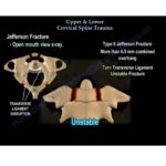
- Jefferson fractures
- Occipitocervical constructs
- Adjacent segment degeneration
- Revision cervical fusion
This allows surgeons to enhance stability without extending fusion levels unnecessarily.
Combined Anterior and Posterior Surgery
Minimally invasive posterior fusion can be combined with anterior cervical procedures in patients with:
- Cervical myelopathy
- Multilevel stenosis
- Cervical kyphosis
- Progressive neurological deficits
Benefits include:
- Improved construct stability
- Smaller posterior incisions
- Reduced muscle injury
- Faster recovery compared with conventional posterior fusion
Stand-Alone Posterior MIS Fusion
Selected elderly patients or those unsuitable for anterior surgery may benefit from stand-alone minimally invasive posterior fusion.
Ideal candidates include patients with:
- Degenerative cervical myelopathy
- Dynamic instability
- Medical comorbidities
- High risk for anterior cervical surgery
Endoscopic decompression combined with facet cage stabilization can provide effective decompression while preserving posterior musculature.
Advantages of Minimally Invasive Posterior Cervical Fusion
Compared with traditional open posterior surgery, minimally invasive techniques offer several advantages:
- Smaller incisions
- Reduced muscle trauma
- Preservation of the nuchal ligament
- Less postoperative pain
- Reduced blood loss
- Lower infection risk
- Shorter hospital stay
- Faster functional recovery
- Improved cosmetic outcomes
Patient Selection
Successful outcomes depend on appropriate patient selection.
Ideal candidates include:
- Cervical pseudarthrosis
- Adjacent segment disease
- Selected cervical fractures
- Cervical instability
- Patients requiring supplemental posterior fixation
- Elderly patients requiring less invasive stabilization
Patients with complex anatomy or severe deformity may still require conventional open surgery.
Surgical Pearls
- Navigation significantly improves implant accuracy.
- Careful preoperative evaluation of vertebral artery anatomy is essential.
- Fluoroscopy remains effective in experienced hands.
- Endoscopic decompression can be combined with minimally invasive fusion techniques.
- Surgeon experience and familiarity with cervical anatomy are critical for optimal outcomes.
Key Takeaways
- Minimally invasive spine surgery emphasizes tissue preservation rather than incision size.
- The anterior cervical approach remains the benchmark for minimally invasive cervical surgery.
- Three posterior minimally invasive fusion techniques are currently available:
- Cervical pedicle screw fixation
- Transfacet screw fixation
- Facet cage placement
- Facet cage technology offers a promising option for selected patients requiring posterior cervical stabilization.
- Combining anterior surgery, endoscopy, and minimally invasive posterior fixation allows surgeons to achieve excellent clinical outcomes while minimizing surgical morbidity.
- Appropriate patient selection and meticulous surgical technique remain essential for successful outcomes.
Leave a Reply