
Courtesy: Prof Nabil Ebraheim, University of Toledo, Ohio, USA
Knee Pain & Meniscal Tears: Clinical Evaluation, Biomechanics, and MRI Interpretation
Core Takeaways
-
Injury Presentation: Meniscal tears typically stem from rotational twisting forces on a weight-bearing knee, manifesting with joint line tenderness, effusion, and mechanical symptoms like locking or clicking.
-
Diagnostic Accuracy: Clinical physical examination alone carries approximately 70% diagnostic accuracy; MRI serves as the definitive non-invasive reference standard.
-
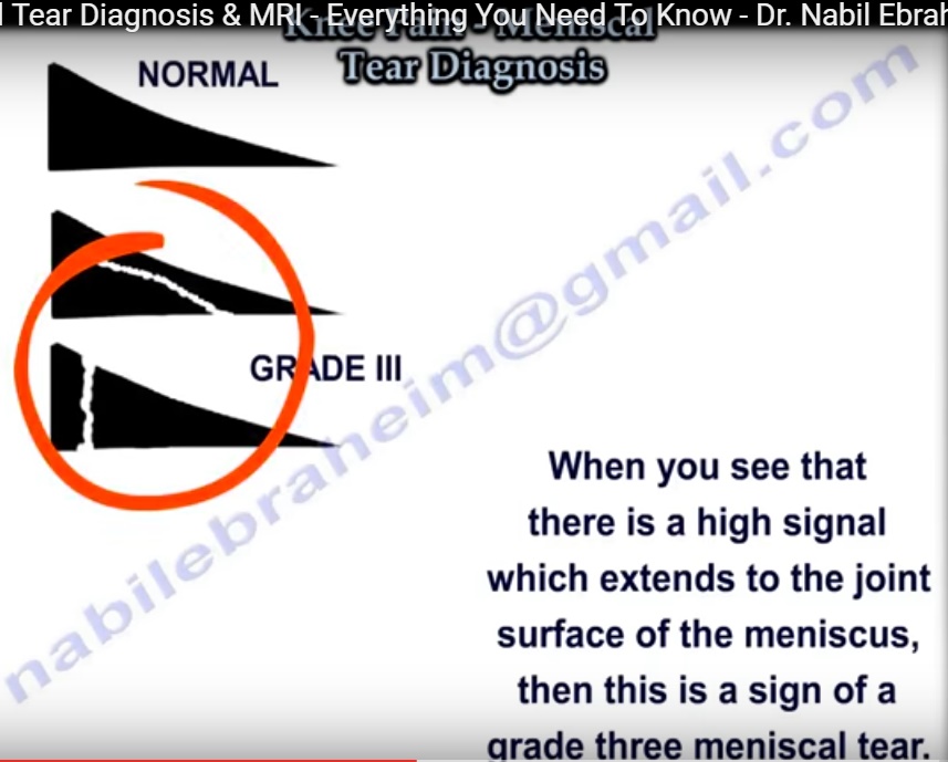
Definitive Tear Criterion: On MRI, a genuine meniscal tear requires Grade 3 hyperintense signal extending completely to an articular surface.
Clinical History & Presentation
-
Mechanism of Injury: Rotational or twisting insult sustained while the knee is loaded or flexed.
-
Mechanical Symptoms:
-
True Joint Locking: Inability to achieve terminal passive or active extension due to a displaced meniscal fragment.
-
Audible or Palpable Clicking: Snapping or clicking sensations during gait or deep flexion.
-
Knee Effusion: Joint swelling, frequently delayed several hours post-injury.
-
Physical Examination Maneuvers
-
Joint Line Tenderness: Localized point tenderness along the medial or lateral joint line.
-
Joint Effusion Assessment: Detection of fluid accumulation (ballottement / fluid wave).
-
McMurray Provocative Test:
-
Technique: Knee is transitioned from full flexion into extension while applying rotational stress to the tibia.
-
Interpretation: A painful palpable or audible click constitutes a positive sign.
-
Localization:
-
Medial Meniscus: Stressed with tibial external rotation.
-
Lateral Meniscus: Stressed with tibial internal rotation.
-
-
Initial Management & Workup Sequence
-
First-Line Conservative Protocol:
-
Oral nonsteroidal anti-inflammatory drugs (NSAIDs).
-
Targeted physiotherapy focusing on range of motion and quadriceps/hamstring conditioning.
-
Intra-articular therapeutic injections when refractory to rest.
-
-
Plain Radiographs (X-rays):
-
Essential initial step prior to advanced imaging.
-
Evaluates underlying bony fractures, degenerative osteoarthritis, or osteochondral lesions.
-
-
Specific Indications for Urgent / Early MRI:
-
True Locked Knee: Inability to extend the knee fully (requires prompt identification of displaced fragments).
-
Aspiration Yielding Hemarthrosis (Bloody Effusion): High clinical suspicion for a concomitant Anterior Cruciate Ligament (ACL) tear, frequently accompanied by an acute meniscal tear.
-
Hidden Pathologies: Most common missed injury on MRI is a vertical tear of the posterior horn of the lateral meniscus, particularly when co-occurring with acute ACL disruptions.
-
Biomechanical Principles & Anatomical Orientation
-
Excursion & Tear Ratios:
-
Lateral Meniscus Excursion: Possesses twice the physiological excursion (mobility) of the medial meniscus.
-
Medial Meniscus Incidence: Due to its rigid peripheral capsular attachment, medial meniscal tears occur three times more frequently than lateral tears.
-
-
Pathology Predilection:
-
Lateral Meniscus: Associated with the vast majority of meniscal cysts and congenital variants.
-
-
MRI Landmark Orientation:
-
Identify the fibula: The lateral meniscus is consistently located on the same side as the fibular head.
-
Spatial location: The fibula is anatomically situated laterally and posteriorly.
-
Normal Meniscal Signal: Pristine, healthy fibrocartilage appears uniformly dark (hypointense / black) on all MRI pulse sequences.
-
Meniscal MRI Grading System
| Grade | MRI Signal Characteristics | Articular Surface Extension | Clinical Significance |
| Normal | Homogeneous, uniform black (hypointense) triangular shape | None | Intact, healthy fibrocartilage |
| Grade 1 | Small, focal, globular area of increased intrasubstance signal | No (confined within substance) | Intrasubstance degeneration; not a tear |
| Grade 2 | Linear, horizontal band of increased intrasubstance signal | No (does not reach surface) | Intermediate degeneration/microtrauma; not a tear |
| Grade 3 | High linear or complex signal intensity | Yes (extends to superior, inferior, or free margin) | True Meniscal Tear |
Morphological Subtypes of Grade 3 Tears
-
Horizontal Cleavage Tears (frequently seen in degenerative joints and associated with meniscal cysts).
-
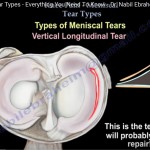
Vertical Tears (longitudinal tears running along the circumferential fibers).
-
Radial Tears (oriented perpendicular to the long axis).
-
Oblique / Flap Tears (“parrot beak” pattern).
-
Complex / Degenerative Tears (multi-directional planes).
-
Displaced / Deficient Fragments (unstable free-floating fragments or flipped segments).
High-Yield MRI Signs on Examinations
-
Double PCL Sign:
-
Visualized on sagittal MRI sequences as an abnormal hypointense band running parallel, anterior, and inferior to the true posterior cruciate ligament (PCL).
-
Caused by a displaced fragment flipped into the intercondylar notch.
-
Pathognomonic: 100% specific for a bucket-handle tear of the medial meniscus.
-
-
Double Anterior Horn Sign:
-
Formed when the posterior horn of the lateral meniscus tears longitudinally and flips anteriorly, seating adjacent to the native anterior horn on sagittal views.
-
Normal Anatomical Variants & MRI False Positives
-
Anterior Transverse Meniscal Ligament:
-
Connects the anterior horn of the medial meniscus to the anterior horn of the lateral meniscus.
-
Enters near the anterior horn of the lateral meniscus, frequently mimicking an anterior horn tear.
-
-
Meniscofemoral Ligaments (Ligament of Humphrey & Ligament of Wrisberg):
-
Arise from the posterior horn of the lateral meniscus and course toward the medial femoral condyle.
-
Intersection with the posterior horn can simulate an acute vertical meniscal tear.
-
-
Popliteus Tendon Hiatus:
-
Normal synovial fluid pooling within the sheath of the popliteus tendon tracks adjacent to the lateral meniscal margin, often falsely suggesting a posterior horn lateral meniscal detachment.
-
Associated Clinical Pathologies
-
Discoid Meniscus:
-
Congenital anatomical variation occurring predominantly on the lateral meniscus.
-
Clinical Presentation: Snapping, clicking, locking, or extension deficits in adolescent/young patients.
-
MRI Diagnostic Criteria: Presence of more than two consecutive “bow-tie” slices on standard 4–5 mm sagittal views, or continuous meniscal tissue extending across more than 50% of the tibial plateau.
-
-
Parameniscal Cyst:
-
Extrusion of intra-articular synovial fluid through a baseline meniscal tear acting as a one-way valve.
-
Predominantly associated with horizontal cleavage tears of the lateral meniscus.
-
Differential Diagnosis: Ganglion cysts, intra-articular loose bodies, or peripheral bursitis.
-
Management: Decompression/aspiration of the cystic cavity combined with arthroscopic partial meniscectomy or tear repair.
-
-
Baker’s (Popliteal) Cyst:
-
Distension of the semimembranosus-medial gastrocnemius bursa in the posterior fossa.
-
Non-specific secondary sign of chronic knee effusion or underlying intra-articular pathology; does not definitively indicate an isolated meniscal tear.
-
Definitive Treatment: Directing therapy or arthroscopic intervention toward repairing/debriding the primary intra-articular trigger (such as a meniscal tear) typically leads to spontaneous resolution of the secondary fluid cyst.
-
Leave a Reply