
Courtesy: Prof Nabil Ebraheim, University of Toledo, Ohio, USA
Low Back Pain, Sciatica, and Spine Differentials: Clinical Reference
Natural History & Conservative Timelines
-
Non-Specific Etiology: Approximately 85% of patients presenting with acute low back pain have no identifiable pathoanatomic cause.
-
Natural Recovery: Roughly 90% of patients recover within 6 weeks of symptom onset.
-
Early Acute Presentation (Under 6 Weeks):
-
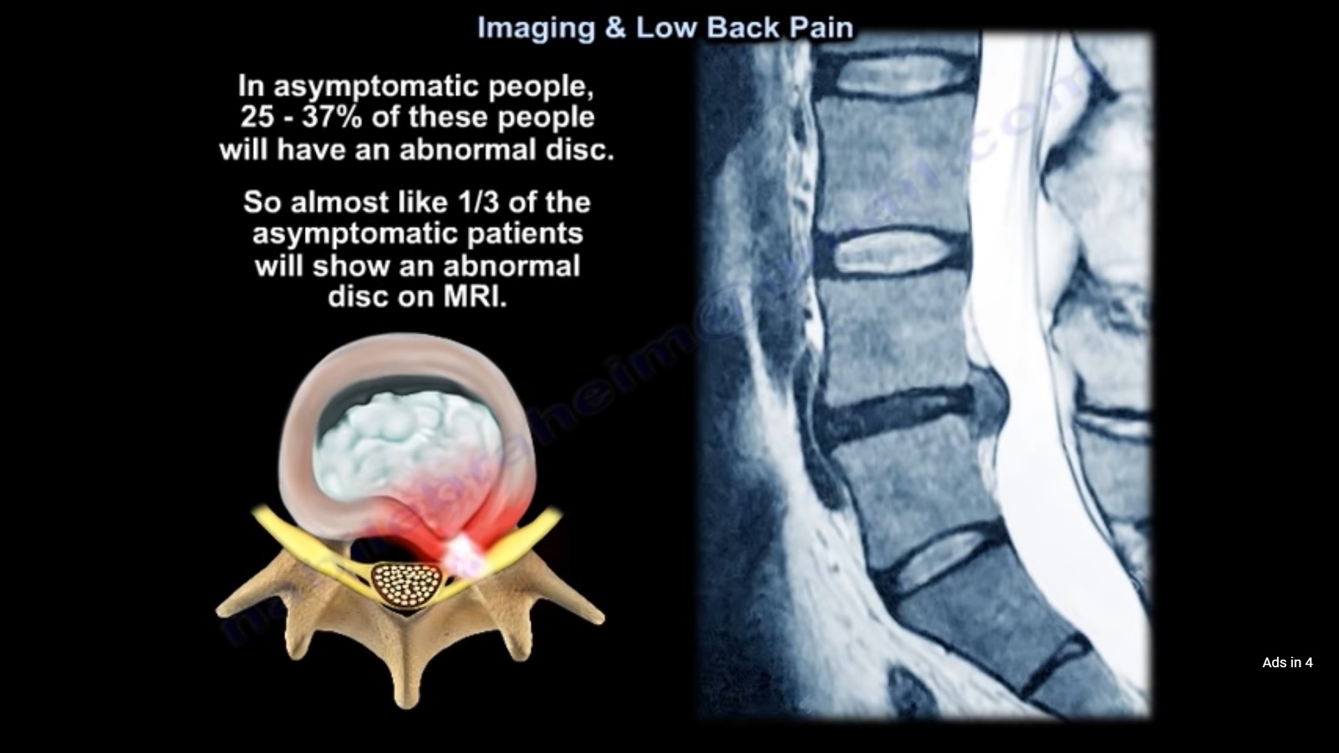
Imaging is not indicated for uncomplicated acute low back pain.
-
Avoid routine plain radiographs, CT scans, and MRI.
-
Management is strictly conservative with activity modification.
-
Patients should return to work early as guided by pain tolerance.
-
-
Indications for Surgical Intervention:
-
Severe, unremitting radicular pain refractory to conservative therapy after 6 to 8 weeks.
-
Progressive or severe neurological motor deficits.
-
Cauda equina syndrome (absolute emergency).
-
-
Chronic Disability Risk: Patients with non-specific low back pain disabled for 6 months without rehabilitation have only a 50% likelihood of ever returning to productive work.
Disc Herniation Anatomy & Neurological Localization
-
Intervertebral Disc Anatomy:
-
Nucleus Pulposus: Central gelatinous core composed predominantly of Type II collagen and hydrophilic proteoglycans.
-
Annulus Fibrosus: Outer concentric lamellar rings composed predominantly of Type I collagen.
-
-
Traversing vs. Exiting Nerve Roots:
-
Paracentral / Posterolateral Herniations: Compress the traversing nerve root in the subarticular lateral recess.
-
Foraminal / Far-Lateral Herniations: Compress the exiting nerve root within or outside the neural foramen.
-
Lumbar Radiculopathy Mapping
-
L4 Nerve Root:
-
Compression etiology: L4–L5 far-lateral (foraminal) disc herniation or L3–L4 posterolateral disc herniation.
-
Motor: Tibialis anterior weakness (impaired ankle dorsiflexion).
-
Reflex: Diminished or absent patellar tendon reflex.
-
Sensory: Medial aspect of the leg and foot.
-
-
L5 Nerve Root:
-
Compression etiology: L4–L5 posterolateral (paracentral) disc herniation.
-
Motor: Extensor hallucis longus (EHL) weakness (impaired great toe extension) and gluteus medius weakness.
-
Gait sign: Trendelenburg gait from hip abductor denervation via the superior gluteal nerve fibers.
-
Reflex: No reliable deep tendon reflex.
-
Sensory: Dorsum of the foot and first web space.
-
-
S1 Nerve Root:
-
Compression etiology: L5–S1 posterolateral disc herniation.
-
Motor: Gastrocnemius-soleus complex and peroneus longus/brevis weakness (impaired plantarflexion).
-
Reflex: Diminished or absent Achilles tendon reflex.
-
Sensory: Lateral border of the foot and plantar aspect of the heel.
-
Urgent Spine Emergencies: Cauda Equina Syndrome
-
Mechanism: Massive central disc herniation occluding the canal and compressing multiple lumbosacral nerve roots.
-
Clinical Red Flags:
-
Bilateral lower extremity radicular pain and progressive motor weakness.
-
Perineal, perianal, and saddle anesthesia.
-
Bladder dysfunction (urinary retention or overflow incontinence) and loss of anal sphincter tone.
-
-
Action: Emergent stat lumbar MRI followed by immediate operative surgical decompression.
Diagnostic Challenges & The Posterior Pelvic Triad
-
The Overlapping Triad: Posterior buttock, groin, and axial pain can arise from three distinct anatomic sources:
-
The lumbar spine.
-
The sacroiliac (SI) joint.
-
The hip joint.
-
-
Differentiating SI Joint Pathology:
-
Physical examination and advanced MRI findings often overlap with spine and hip pathology.
-
The gold standard diagnostic modality is a fluoroscopically guided diagnostic intra-articular SI joint injection using local anesthetic.
-
-
Post-Spine Arthrodesis Hip Pathology:
-
Persistent anterior groin and anterior thigh pain following lumbar spine surgery is a hallmark of occult hip osteoarthritis.
-
True hip pathology characteristically refers to the anterior groin and proximal thigh, not the posterior buttock.
-
Neurogenic vs. Vascular Claudication & Diagnostic Pearls
-
Vascular Screening: Palpation of distal peripheral pulses (dorsalis pedis and posterior tibial) is mandatory in all patients presenting with neurogenic claudication to rule out coexisting peripheral artery disease.
-
Physical Provocative Signs:
-
Straight Leg Raise (SLR) Test: The most sensitive dynamic physical tension test for acute lumbar disc herniations.
-
Pain exacerbated by seated posture reflects increased intradiscal pressure and sciatic nerve tension.
-
-
Stationary Bicycle Test:
-
Patients with lumbar spinal stenosis can cycle pain-free because trunk flexion widens the spinal canal.
-
Patients with vascular claudication develop worsening ischemic leg cramps while cycling due to muscular metabolic demand.
-
-
Upper Motor Neuron Red Flags:
-
Low back pain accompanied by hyperreflexia, clonus, or spastic gait disturbance represents an upper motor neuron lesion.
-
This cannot originate from the lumbar canal; it requires an immediate cervical spine MRI to evaluate for cervical spondylotic myelopathy.
-
-
Spine Oncology & Infection Signs:
-
Infection (Diskitis/Osteomyelitis): Typically crosses and destroys the intervertebral disc space.
-
Metastatic Malignancy: The spine is the most frequent site of bony metastasis. Tumors seed the vascular vertebral body and erode the pedicle, producing the winking owl sign (absent pedicle) on AP radiographs while sparing the disc space.
-

Leave a Reply