
Courtesy: Prof Nabil Ebraheim, University of Toledo, Ohio, USA
Spondylolysis, Spondylolisthesis, Spondylitis, and Spondylosis: Diagnostic Differentiation and Clinical Protocols
Terminology Matrix & Structural Distinctions
-
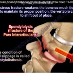
Spondylolysis: Structural bone defect, discontinuity, or non-union involving the pars interarticularis of the vertebral neural arch.
-
Spondylolisthesis: Pathological translational slippage of one vertebral body relative to the subjacent vertebra (not isolated displacement of the intervertebral disc).
-
Spondylitis: Primary inflammatory or infectious condition affecting the vertebral bodies, endplates, and associated axial ligaments (e.g., Ankylosing Spondylitis, tuberculous spondylodiscitis).
-
Spondylosis: Age-related degenerative osteoarthropathy of the spinal motion segment (involving intervertebral discs, uncovertebral joints, and facet articulations).
1. Spondylolysis
-
Epidemiology & Location:
-
Affects approximately 5% of the general population.
-
Predominantly localized to the L5 vertebra (accounting for roughly 85% to 90% of cases).
-
-
Biomechanical Provocation: Repetitive lumbar hyperextension and rotation (common in gymnasts, fast bowlers, divers, and offensive linemen) produce cyclic fatigue stresses across the pars.
-
Diagnostic Workup:
-
Oblique Plain Radiographs: Classic demonstration of the Scotty Dog with a collar sign, where the defect represents a lucent fracture line traversing the neck of the dog (pars interarticularis).
-
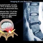
Advanced Imaging: If plain radiographs are negative in an active adolescent with focal extension back pain, obtain a SPECT bone scan or thin-slice cross-sectional CT scan / MRI to detect early stress reactions or non-displaced fatigue fractures.
-
2. Spondylolisthesis
-
Age and Demographics:
-
Pediatric / Adolescent Cohorts: Most frequently isthmic or dysplastic spondylolisthesis occurring at L5–S1.
-
Adult Cohorts: Most frequently degenerative spondylolisthesis occurring at L4–L5, with a strong female predominance.
-
-
Neurological Localization:
-
In adult degenerative L4–L5 slips, anterior translation typically compresses the traversing L5 nerve root within the lateral recess.
-
In high-grade or symptomatic pediatric L5–S1 isthmic slips, the L5 nerve root is classically impinged beneath the fibrocartilaginous pseudarthrosis callus of the mobile posterior arch (Gill body) or within the exiting foramen.
-
-
Slip Progression Dynamics:
-
Approximately 15% of patients with an isolated pars defect progress to forward translational slippage.
-
In adult degenerative slips, translation rarely exceeds 30% (Meyerding Grade I or low Grade II) due to stabilization by intact posterior elements and competent facet capsular tethers.
-
Dysplastic (congenital) slips and high-grade isthmic slips in skeletally immature patients carry the highest risk of unchecked mechanical progression.
-
3. Spondylitis
-
Natural History (Ankylosing Spondylitis): Progresses systematically from an active inflammatory phase (sacroiliitis, enthesitis) to chronic tissue ossification, resulting in widespread fusion and the rigid “bamboo spine”.
-
Radiographic Signatures:
-
Fine, vertically oriented marginal syndesmophytes bridging contiguous vertebral endplates.
-
Progressive squaring of vertebral bodies and complete ankylosis of the sacroiliac and facet joints.
-
-
The Fragile Ankylosed Spine & Occult Trauma:
-
The fused spine behaves biomechanically as a long, brittle, osteopenic lever arm.
-
Minor ground-level falls can trigger catastrophic chalk-stick fractures, most commonly localized to the lower cervical spine or cervicothoracic junction.
-
Fractures are frequently non-displaced and occult on plain radiography, mandating immediate CT or MRI evaluation.
-
High risk of acute epidural hematoma and progressive neurological deficit.
-
Surgical Protocol: Requires prompt decompression (laminectomy) combined with long-segment rigid posterior instrumented fusion to control long-lever-arm instability.
-
4. Spondylosis: Stenosis, Radiculopathy, and Cervical Myelopathy
-
Pathophysiology: Facet joint hypertrophy, uncovertebral osteophytosis, and disc collapse cause neuroforaminal stenosis (producing radiculopathy) or spinal canal stenosis (producing myelopathy or neurogenic claudication).
-
Cervical Spondylotic Myelopathy (CSM) Manifestations:
-
Gait Instability: Characterized by a slow, wide-based, clumsy, or shuffling gait.
-
Upper Extremity Dysmetria: Loss of fine motor coordination (difficulty buttoning shirts, manipulating keys, deterioration of handwriting).
-
The Myelopathic Hand: Intrinsic hand muscle wasting (particularly dorsal interossei) and positive finger escape sign.
-
-
Upper Motor Neuron (UMN) Exam Signs:
-
Hoffman Sign: Involuntary flexion/adduction of the thumb and index finger upon quick flicking of the middle fingernail.
-
Babinski Sign / Extensor Plantar Response: Dorsiflexion (extension) of the great toe with fanning of the lesser toes upon lateral plantar stroke.
-
Pathological hyperreflexia and sustained ankle clonus.
-
Tandem Spinal Stenosis: Diagnostic Trap Protocol
-
The Exam & Clinical Pitfall:
-
An elderly patient presents with progressive walking difficulty, gait imbalance, and leg weakness.
-
Lumbar spine MRI demonstrates high-grade lumbar spinal canal stenosis.
-
Physical examination reveals lower-extremity hyperreflexia, sustained clonus, a positive Babinski sign, or a positive Hoffman sign.
-
-
Clinical Decision Rule:
-
Lumbar spinal stenosis causes lower motor neuron (LMN) pathology (hyporeflexia, flaccidity) or neurogenic claudication. It never produces upper motor neuron signs.
-
The presence of UMN signs (hyperreflexia, extensor plantar response) proves the coexistence of proximal cord compression (Tandem Spinal Stenosis).
-
-
Next Diagnostic Step:
-
Obtain an immediate cervical spine MRI (or CT myelogram if MRI is contraindicated due to non-compatible implants or severe claustrophobia).
-
Prioritize decompression of the symptomatic cervical myelopathy prior to addressing the lumbar spine, as cervical cord decompression frequently improves the patient’s functional stability and reduces anesthesia-related hyperextension risks.
-


Leave a Reply