
Courtesy: Prof Nabil Ebraheim, University of Toledo, Ohio, USA
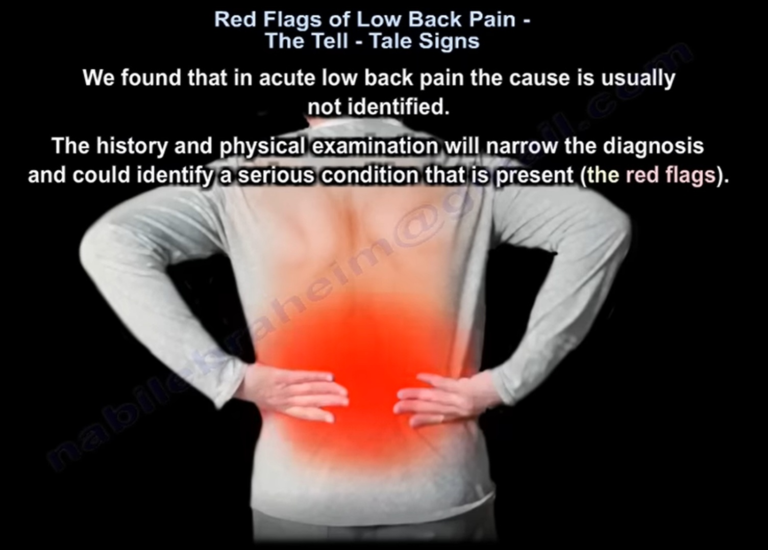
Lumbar Disc Herniation: Pathoanatomy, Neurological Localization, and Management
Structure and Mechanical Function of the Intervertebral Disc
-
Anatomical Definition: A fibrocartilaginous cushion interposed between adjacent vertebral bodies that absorbs axial shock and permits multidirectional spinal motion.
-
Component Anatomy:
-
Nucleus Pulposus: Inner hydrated, gelatinous core predominantly composed of water, proteoglycans, and Type II collagen.
-
Annulus Fibrosus: Outer concentric lamellar rings predominantly composed of tensile Type I collagen that contain the nuclear core.
-
-
Mechanism of Herniation: Tearing or attenuation of the annulus fibrosus allows displacement of nucleus pulposus tissue beyond the physiological intervertebral disc boundaries.
Anatomical Vectors of Disc Displacement
-
Posterolateral (Paracentral) Herniation:
-
The most common presentation.
-
Displaces into the lateral recess and compresses the traversing nerve root.
-
Example: An L4–L5 posterolateral disc herniation impinges the traversing L5 nerve root.
-
-
Foraminal / Far-Lateral Herniation:
-
Represents approximately 10% of lumbar disc herniations.
-
Displaces directly within or lateral to the neural foramen, compressing the exiting nerve root.
-
Example: An L4–L5 far-lateral disc herniation impinges the exiting L4 nerve root.
-
-
Central Herniation (The Cauda Equina Emergency):
-
Massive midline extrusion directly occluding the central thecal sac.
-
Compresses multiple descending lumbosacral nerve roots of the cauda equina.
-
Cauda Equina Syndrome: Presentation and Surgical Window
-
Clinical Triad & Red Flags:
-
Severe axial low back pain with bilateral lower-extremity sciatica or weakness.
-
Saddle and perineal anesthesia (loss of sensation in the S3–S5 distribution).
-
Bladder dysfunction (urinary retention or overflow incontinence) and loss of voluntary anal sphincter tone.
-
-
Management Protocol:
-
Obtain an immediate emergency contrast or non-contrast lumbar MRI.
-
Proceed with emergent surgical decompression within 48 hours of onset to maximize the likelihood of bladder, bowel, and motor recovery.
-
Sciatica and Neurological Radiculopathy
-
Etiology: Mechanical compression and chemical irritation of lumbosacral nerve roots (typically L5 or S1) from herniated disc fragments, or peripheral entrapment along the sciatic nerve trunk (e.g., piriformis syndrome).
-
Clinical Characteristics:
-
Sharp, shooting, electric pain originating in the lower back and buttock, radiating down the leg into the foot.
-
Unilateral burning, tingling, and paresthesias.
-
Pain is exacerbated by sitting (increased intradiscal pressure and nerve tension) and alleviated by standing or recumbent positioning.
-
-
Physical Tension Sign (Straight Leg Raise / SLR):
-
Performed by passively raising the extended lower extremity.
-
A positive test reproduces radiating leg pain and paresthesia between 30° and 70° as the sciatic nerve stretches across the disc protrusion. Isolated back pain does not constitute a positive test.
-
-
Dermatomal, Motor, and Reflex Distribution:
-
L4 Root: Medial leg/foot sensation; tibialis anterior motor weakness (impaired ankle dorsiflexion); diminished patellar tendon reflex.
-
L5 Root: Dorsum of the foot and first web space sensation; extensor hallucis longus motor weakness (impaired great toe extension); no reliable tendon reflex.
-
S1 Root: Lateral border and sole of the foot sensation; gastrocnemius-soleus motor weakness (impaired plantarflexion); diminished Achilles tendon reflex.
-
Conservative Management vs. Surgical Predictors
-
Conservative Window: Initiate at least 6 weeks of structured conservative treatment (analgesics, anti-inflammatory medications, physical therapy, and activity modification) before considering elective surgery.
-
Spontaneous Resorption: Sequestered and extruded disc herniations carry the highest biological rate of spontaneous macrophage-mediated enzymatic resorption.
-
Predictors of Successful Surgical Outcome:
-
Leg pain exceeding axial low back pain.
-
A concordant positive Straight Leg Raise test.
-
Objective neurological deficits (dermatomal sensory loss, motor weakness, or reflex changes).
-
Exact anatomical correlation between physical exam findings and cross-sectional MRI imaging.
-
Related Posts
 Lumbar Disc Herniation
Part 1 Part 2 Courtesy: Courtesy: Douglas Gillard, BS, DC, Spine Researcher http://chirogeek.com/
 Acute Low Back Ache and Lumbar Disc Herniation
Courtesy: Prof Nabil Ebraheim, University of Toledo, Ohio, USA
- Lumbar disc Herniation Basics
Courtesy : Prof Nabile Ebraheim, Chairman of Orthopaedic Surgery, University of Toledo, Ohio, United states
Leave a Reply