
Courtesy: Prof Nabil Ebraheim, University of Toledo, Ohio, USA
Acute Low Back Pain and Sciatica: Natural History, Red Flags, and Differential Diagnosis
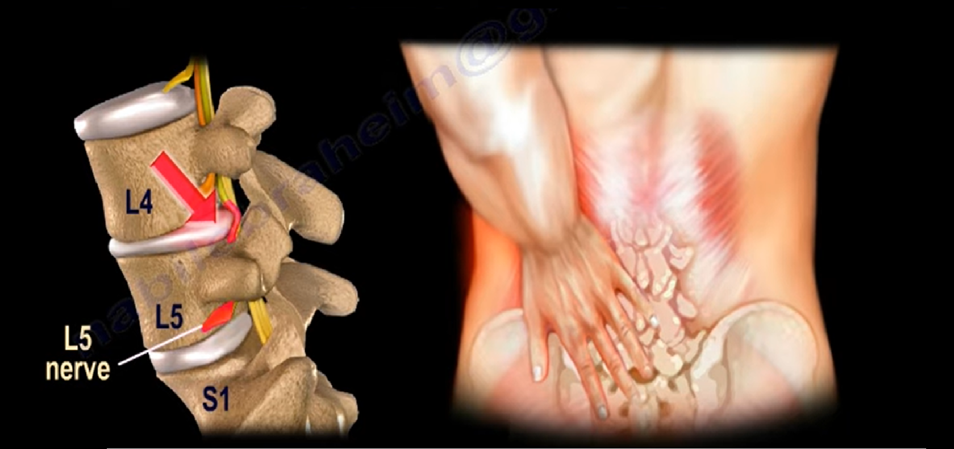
Natural History & Biomechanics of the Intervertebral Disc
-
Initial Conservative Window: Both acute low back pain and sciatica require an initial 6-week trial of conservative therapy.
-
Treatment Regimen: Physical therapy, anti-inflammatory medications (NSAIDs), and symptom-guided activity modification.
-
Imaging Threshold: Even when an MRI demonstrates a large herniated disc, continue conservative management for 6 weeks unless progressive neurological deficits or cauda equina syndrome are present.
-
Spontaneous Recovery: Approximately 90% of patients experience resolution of acute symptoms within 1 month.
-
Risk Factors for Chronic Low Back Pain: Cigarette smoking, clinical depression, and occupational exposure to whole-body vibration.
-
Intradiscal Pressure Variations (Nachemson Biomechanics):
-
Lowest Intradiscal Pressure: Supine position (approximately 25% of standing baseline).
-
Baseline Intradiscal Pressure: Upright standing (100%).
-
Intermediate Pressure: Upright seated posture (140%).
-
Highest Intradiscal Pressure: Seated, leaning forward, and holding a weight (reaches up to 275% of standing baseline).
-
Spinal Oncology & Metastatic Disease
-
Clinical Presentation: Pain that persists at rest and worsens at night is a critical red flag for spinal neoplasm.
-
Prior Malignancy History: Any history of cancer presenting with back pain mandates immediate plain radiography and contrast-enhanced MRI.
-
Renal Cell Carcinoma: Hypervascular spinal metastases from renal tumors require preoperative spinal angiography and transarterial embolization to control intraoperative hemorrhage.
-
Anatomical Route of Metastasis: Hematogenous seeding occurs primarily in the posterior vertebral body, with secondary extension into the pedicle.
-
Radiographic Detection Threshold: Between 30% and 40% of trabecular bone mass must be resorbed before a lytic lesion becomes visible on plain radiographs.
-
The Winking Owl Sign: Unilateral lytic destruction of a pedicle on an AP radiograph causes the appearance of an owl winking.
Spinal Infection (Spondylodiscitis)
-
Anatomical Target: Bacterial infection primarily involves and destroys the intervertebral disc space and adjacent vertebral endplates.
-
Laboratory Markers: Erythrocyte sedimentation rate (ESR) and C-reactive protein (CRP) are consistently elevated.
-
Diagnostic Sensitivity:
-
Fever is present in only 50% of patients with spondylodiscitis.
-
Leukocytosis (elevated WBC count) is present in less than 50% of patients.
-
Blood cultures are positive in approximately 24% of cases.
-
-
Diagnostic Strategy: Obtain an urgent MRI and perform an image-guided biopsy to direct targeted culture-specific antibiotic therapy.
-
Surgical Indications: Presence of an epidural abscess, spinal instability, or progressive neurological deterioration requires prompt surgical decompression.
-
Postoperative Infection Monitoring: Serum CRP is the most sensitive marker to detect early postoperative infection because it normalizes much faster than ESR.
Osteoporotic Vertebral Compression Fractures
-
The Osteoporotic Cascade: Osteoporotic fragility fractures classically follow a predictable anatomical sequence:
-
First: Distal radius (Colles fracture).
-
Second: Thoracolumbar spine (vertebral compression fracture).
-
Third: Proximal femur (hip fracture).
-
-
Cascade Effect: The presence of a single vertebral compression fracture significantly increases the risk of subsequent vertebral fractures.
-
Medical Prevention:
-
Medical management (e.g., antiresorptive agents) reduces the incidence of secondary vertebral fractures by 60% after 1 year of treatment.
-
Medical management reduces secondary vertebral fractures by 40% at 2 years.
-
-
Steroid Red Flag: History of prolonged corticosteroid use warrants early plain radiography to evaluate for occult insufficiency fractures.
Seronegative Spondyloarthropathy vs. DISH
-
Ankylosing Spondylitis (AS):
-
Originates at the sacroiliac (SI) joints with bilateral, symmetric sacroiliitis.
-
Strongly associated with HLA-B27 positivity.
-
Displays marginal syndesmophytes (thin, vertical calcifications) with diffuse ossification of the outer annulus fibrosus and intervertebral disc spaces.
-
Lacks large, bulky, protruding osteophytes.
-
-
Diffuse Idiopathic Skeletal Hyperostosis (DISH):
-
Strongly associated with type 2 diabetes mellitus, advanced age, and metabolic syndrome; screen with Hemoglobin A1c.
-
Displays non-marginal syndesmophytes and bulky, coarse osteophytes.
-
Characterized by confluent, flowing ossification along the anterolateral aspect of at least four contiguous vertebral bodies.
-
Sacroiliac joints and intervertebral disc heights are characteristically preserved.
-
Advanced MRI Diagnostics: Contrast Differentiation
-
Asymptomatic False-Positive MRI Rates:
-
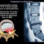
Up to 35% of asymptomatic individuals under 40 years of age demonstrate disc abnormalities or protrusions on routine MRI.
-
Up to 90% of asymptomatic individuals over 60 years of age have positive findings of disc degeneration or canal stenosis.
-
-
Gadolinium-Enhanced MRI (Recurrent Disc vs. Peridural Scar):
-
Routine T1 and T2 non-contrast sequences cannot reliably differentiate recurrent disc herniation from post-surgical epidural granulation scar tissue.
-
Epidural Granulation Scar Tissue: Vascularized tissue that rapidly absorbs gadolinium, demonstrating early, robust contrast enhancement. Treat non-operatively.
-
Recurrent Disc Herniation: Avascular, necrotic nuclear material that fails to enhance with gadolinium contrast. Symptomatic recurrent discs causing persistent radiculopathy may require revision discectomy.
-
Leave a Reply