Courtesy: Prof Nabil Ebraheim, University of Toledo, Ohio, USA
Sciatic Nerve Anatomy
- Sciatic nerve is the largest nerve in the body.
- Originates from L4, L5, S1, S2, and S3 nerve roots.
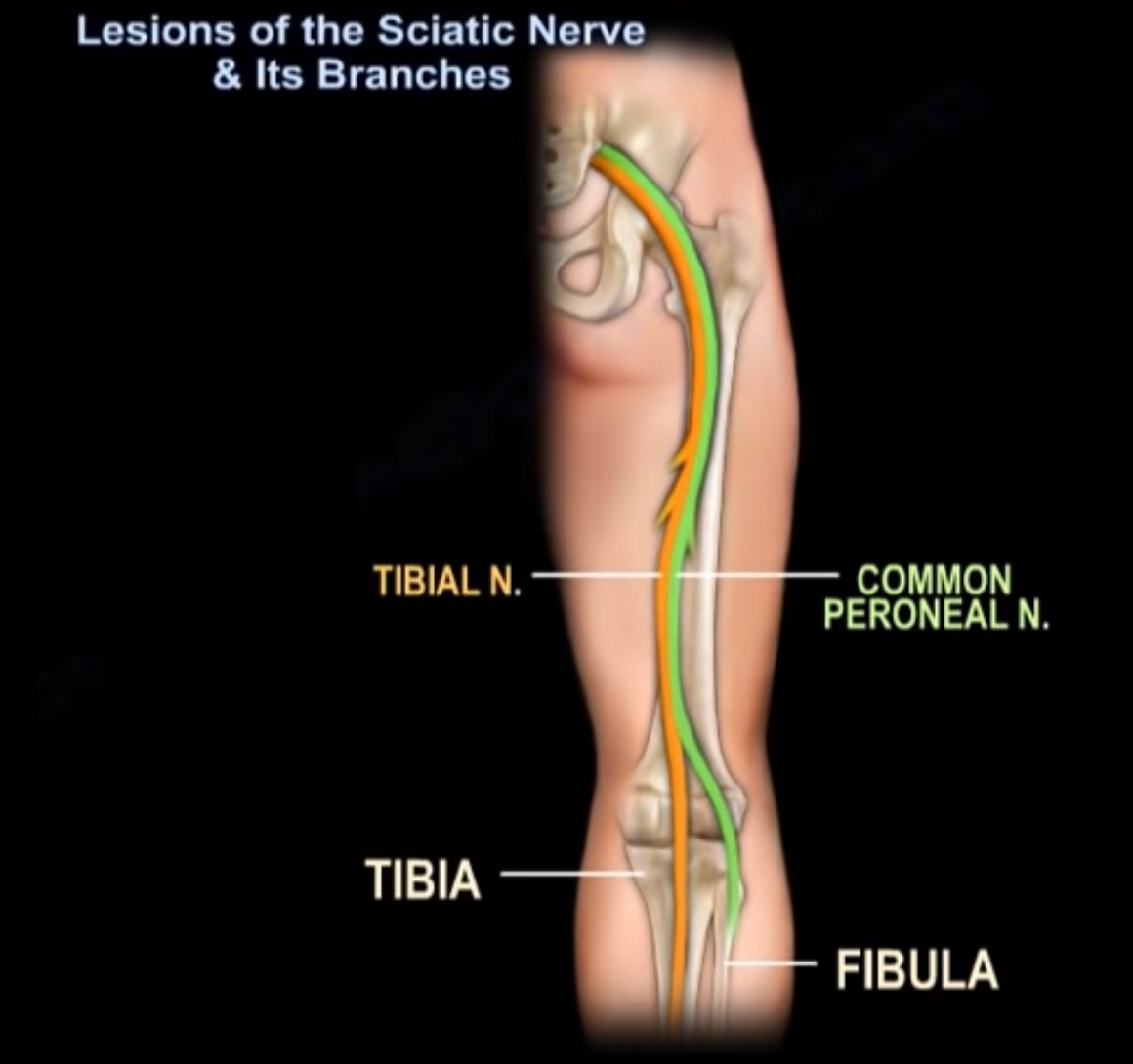
- Contains two major components: tibial nerve and common peroneal (fibular) nerve.
- Travels through gluteal region and posterior thigh.
- Usually divides into tibial and common peroneal nerves in the distal thigh.
- In about 10% of individuals division occurs at the greater sciatic foramen.
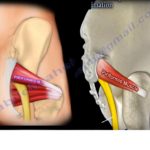
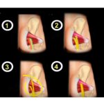
Relationship with Piriformis Muscle
- Sciatic nerve normally exits pelvis inferior to piriformis muscle.
- Some anatomical variations may exist.
- Compression of sciatic nerve at piriformis causes piriformis syndrome.
Piriformis Syndrome
- Sciatic nerve compression by piriformis muscle.
- Causes buttock pain and sciatica-like symptoms.
- Must rule out lumbar disc herniation before diagnosis.
- Diagnosis is clinical and by exclusion.
Clinical Tests for Piriformis Syndrome
- Lasègue maneuver: hip flexion to 90° with knee extended reproduces pain.
- FAIR test: Flexion, Adduction, and Internal Rotation of hip stretches piriformis.
- Pressure over buttock may reproduce tenderness.
Course of Sciatic Nerve in Gluteal Region
- Nerve runs anterior to piriformis muscle.
- Posterior to obturator internus, gemelli, and quadratus femoris.
- Lies between greater trochanter and ischial tuberosity.
Sciatic Nerve Injuries
- Commonly injured in posterior hip dislocation.
- Common peroneal division most frequently affected.
- Can be injured during posterior hip surgery or acetabular screw placement.
- Traction during surgery should be applied with knee flexed to reduce nerve tension.
- Injury may cause foot drop.
Muscles Supplied by Sciatic Nerve
- Long head of biceps femoris.
- Ischial portion of adductor magnus.
- Short head of biceps femoris supplied by common peroneal division.
Common Peroneal Nerve
- Branches from sciatic nerve near the knee.
- Wraps around the neck of the fibula.
- Divides into superficial and deep peroneal nerves.
- Vulnerable to injury in knee dislocation and fibular neck fractures.
Superficial Peroneal Nerve
- Supplies lateral compartment muscles: peroneus longus and peroneus brevis.
- Pierces fascia in distal third of leg to become superficial.
- Provides sensation to dorsum of the foot.
Injury to Superficial Peroneal Nerve
- May occur during lateral compartment fasciotomy.
- Risk during anterolateral extensile approach for pilon fractures.
- May be injured during ankle arthroscopy portal placement.
Deep Peroneal Nerve
- Supplies anterior compartment muscles: tibialis anterior, extensor hallucis longus, extensor digitorum longus.
- Provides motor supply to extensor digitorum brevis.
- Provides sensory innervation to the first web space.
Deep Peroneal Nerve Injury
- Causes foot drop due to loss of ankle dorsiflexion.
- Loss of sensation in the first web space.
Anterior Tarsal Tunnel Syndrome
- Compression of deep peroneal nerve beneath inferior extensor retinaculum.
- Symptoms include dorsal foot pain, paresthesia, and numbness in first web space.
- Symptoms worsen with tight shoes and plantar flexion.
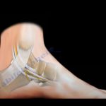
Tibial Nerve
- Runs posterior to medial malleolus beneath flexor retinaculum.
- Divides into medial plantar, lateral plantar, and medial calcaneal branches.
Tarsal Tunnel
- Fibro-osseous tunnel behind medial malleolus.
- Covered by flexor retinaculum.
- Contains structures remembered by mnemonic: ‘Tom, Dick, And Very Nervous Harry’.
- Tibialis posterior tendon.
- Flexor digitorum longus tendon.
- Posterior tibial artery.
- Tibial nerve.
- Flexor hallucis longus tendon.
Tarsal Tunnel Syndrome
- Compression neuropathy of posterior tibial nerve within the tarsal tunnel.
- Causes plantar foot pain, numbness, and paresthesia.
Baxter’s Nerve
- First branch of lateral plantar nerve.
- Provides motor supply to abductor digiti minimi.
- Responsible for up to 20% of heel pain cases.
- Entrapment may mimic plantar fasciitis.
Sites of Baxter’s Nerve Entrapment
- Between fascia of abductor hallucis and quadratus plantae muscle.
- Near medial calcaneal tuberosity.
Calcaneal Traction Pin
- Used for traction or external fixation.
- Inserted from medial to lateral side of calcaneus.
- Pin should be placed posteroinferior to avoid neurovascular bundle.
Morton’s Neuroma
- Compression neuropathy of interdigital nerve.
- Most commonly occurs in third interdigital space.
- Associated with perineural fibrosis.
Leave a Reply