Courtesy: Prof Nabil Ebraheim, University of Toledo, Ohio, USA
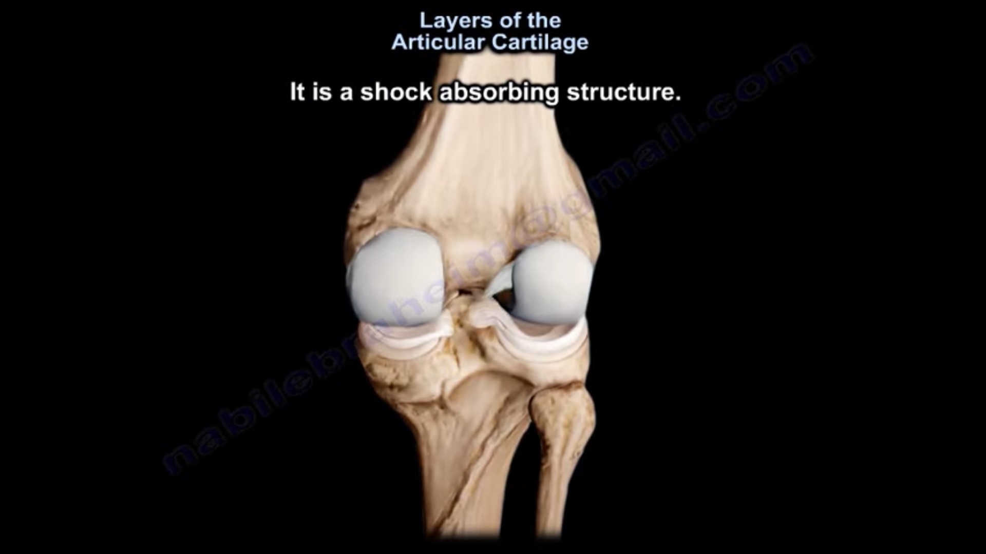
Layers of Articular Cartilage
- The articular cartilage is avascular, meaning that it does not have a blood supply and does not have nerves.
- It is a shock absorbing structure.
- Bone is made first, so bone contains collagen Type I.
- Cartilage is made after the bone, so cartilage contains collagen Type Il.
In this lecture, we will highlight three things:
1.Composition of the cartilage.
2.Layers of the cartilage.
3If the cartilage is injured, can it heal?
Cartilage is made of water, collagen Type II and proteoglycans.
- Water (about 74%)
- Collagen (about 15%)
- Proteoglycans (about 1%)
- Other material (less than 1%)
- The cartilage needs water for lubrication and for transport of nutrients.
- The cartilage will need collagen to give the cartilage the tensile strength and stiffness.
- The collagen makes a mesh work that is both flexible and tough, but entraps the proteoglycans and the cartilage cells.
- The cartilage cells (chondrocytes) is about 1% and it is responsible for the synthesis, the maintenance, and the hemostasis of cartilage.
SOX9 is the master switch for differentiation of cells of chondrocyte lineage.
- The other important structure is the proteoglycan.
ProteoGlycan Vs GlycosaminoGlycan
- The cartilage proteoglycans are large, long chains of negative charged molecules.
- The glycosaminoglycans are linear polysaccharides such as keratin sulfate and chondroitin sulfate
- The proteoglycan is a core protein
- glycosaminoglycan chains: consist of chondroitin sulfate and the keratin sulfate, and this is called proteoglycan monomer.
Chondroitin sulfate is the most prevalent glycosaminoglycan
- It becomes a proteoglycan aggregate when it gets connected to the hyaluronic acid by a link protein.
- The cartilage proteoglycan is multiple glycosaminoglycans bound to a core protein which is bound to hyaluronic acid through a link protein.
The link protein connects the proteoglycan monomer to the hyaluronic acid backbone.
- Proteoglycans can be associated with up to 50% of its weight in water (the proteoglycan swells).
- It is responsible for the swelling pressure of the cartilage and the collagen restrains that swelling.
- The proteoglycans attract water because of the long chains of negative charges
- That will increase the swelling and that fluid pressure provides strength in compression.
- The swelling pressure in the cartilage is predominantly due to the association of its changeable water with the aggrecan.
Aggrecan is associated with 50 times its weight in water.
- The aggrecan aggregates on hyaluranic acid with a link protein.
- It has a longer core protein with multiple keratin sulfate and chondroitin sulfate chains.
Leave a Reply