Prof Nabil Ebraheim, University of Toledo, Ohio, USA
Knee pain is a very common complaint and may arise from:
-
The knee cap (patella) itself
-
The tendons attached to the patella (patellar tendon and quadriceps tendon)
-
Structures inside or around the knee joint such as menisci, ligaments, bursae, or cartilage
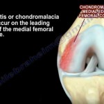
1. Chondromalacia Patella (Patellofemoral Pain Syndrome)
-
One of the most common causes of anterior knee pain
-
Caused by softening and degeneration of the cartilage on the underside of the patella
-
Cartilage may show:
-
Erosions
-
Defects
-
Partial or full-thickness damage
-
-
Pain is felt in the front of the knee
-
More common in young individuals
-
Pain worsens with:
-
Climbing stairs
-
Going down stairs
-
Prolonged sitting
-
Treatment
-
Non-steroidal anti-inflammatory drugs (NSAIDs)
-
Physiotherapy
-
Surgery is rarely required
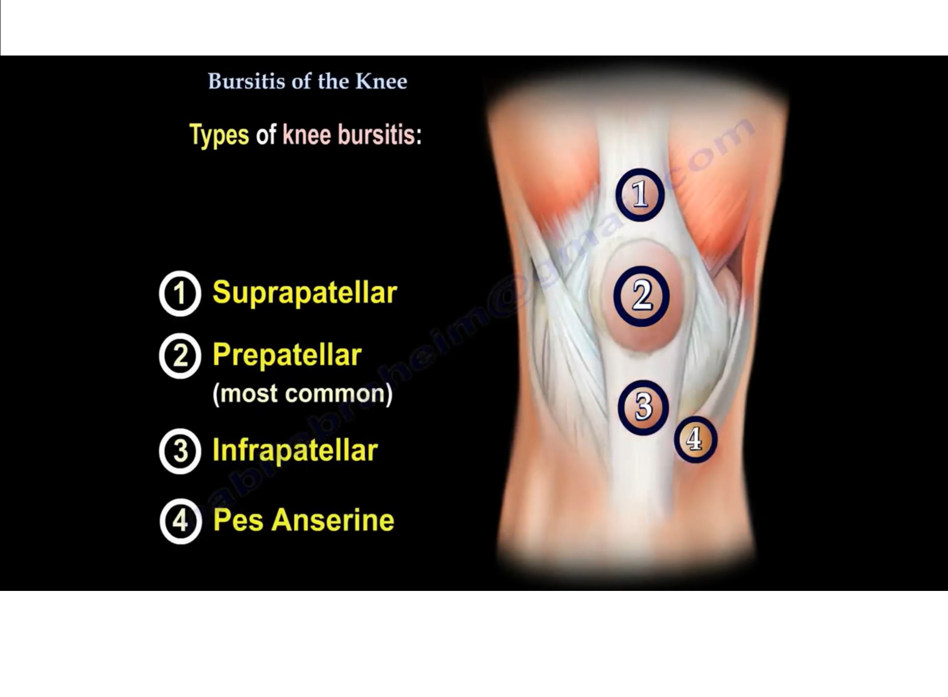
2. Prepatellar Bursitis
-
Pain and swelling occur directly in front of the knee cap
-
Caused by inflammation of the prepatellar bursa
-
The bursa fills with fluid, leading to:
-
Swelling
-
Tenderness
-
Localized lump over the patella
-
-
Pain in front of the knee above or below the patella may also suggest associated tendonitis
3. Patellar Tendonitis (Jumper’s Knee)
-
The patellar tendon works together with the quadriceps tendon to straighten the knee
-
An overuse injury, commonly seen in athletes involved in jumping sports
-
Accounts for approximately 20% of jumping-related knee injuries
-
Pain is:
-
Activity-related
-
Located below the knee cap
-
-
Tenderness is present at the distal pole of the patella
-
Tender in knee extension
-
Less tender in knee flexion
-
-
Predisposing factors:
-
Quadriceps tightness or weakness
-
Hamstring tightness
-
Muscle imbalance or atrophy
-
Treatment
-
Anti-inflammatory medications
-
Stretching and strengthening exercises
-
Hamstring and quadriceps stretching
-
Eccentric strengthening programs
-
Most early cases respond well to non-operative treatment
-
Surgery (tendon debridement and repair):
-
Reserved for severe cases
-
Considered if symptoms persist after 6–12 months of conservative treatment
-
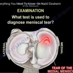
4. Meniscal Tear
-
The meniscus acts as a shock absorber, protecting knee cartilage
-
Injury causes pain on the:
-
Medial (inner) or
-
Lateral (outer) joint line
-
-
Common symptoms include:
-
Joint line pain
-
Swelling
-
Locking or catching sensation
-
Feeling of instability
-
-
Joint line tenderness is the most important clinical finding
Clinical Test
-
McMurray’s test
-
Painful click or pop during knee movement from flexion to extension with rotation
-
Imaging
-
MRI is very helpful for diagnosis
5. Knee Arthritis (Osteoarthritis)
-
A very common condition, especially with increasing age
-
Cartilage cells gradually degenerate and repair capacity decreases
-
Leads to:
-
Progressive cartilage loss
-
Joint space narrowing
-
Bony changes visible on X-rays
-
-
Symptoms include:
-
Pain
-
Stiffness
-
Reduced mobility
-
6. Baker’s Cyst (Popliteal Cyst)
-
A fluid-filled swelling located behind the knee
-
Filled with synovial fluid
-
Located between:
-
Semimembranosus tendon
-
Medial head of gastrocnemius
-
-
Often associated with:
-
Arthritis
-
Meniscal tears
-
7. Ligament Injuries of the Knee
-
Commonly occur due to sports-related trauma
Medial Collateral Ligament (MCL) Injury
-
Most commonly injured knee ligament
-
Involves the ligament on the inner side of the knee
Anterior Cruciate Ligament (ACL) Injury
-
Often caused by valgus stress and twisting injury
-
Common features:
-
Sudden swelling
-
Hematoma
-
-
Lachman’s test is usually positive
-
MRI confirms the diagnosis
8. Iliotibial Band Syndrome (ITBS)
-
Caused by inflammation and thickening of the iliotibial band
-
Results from repetitive friction over the lateral femoral condyle
-
The IT band:
-
Extends from the iliac crest to the knee
-
Slides back and forth during knee movement
-
-
Maximum impingement occurs around 30° of knee flexion
Clinical Features
-
Pain on the outer side of the knee
-
Swelling, tenderness, and crepitus over the lateral femoral condyle
-
Common in:
-
Runners
-
Cyclists
-
Athletes with repetitive knee flexion-extension
-
-
Pain may be reproduced with a single-leg squat
-
Ober’s test assesses IT band tightness
Imaging
-
MRI may show edema along the IT band
Treatment
-
Primarily non-operative:
-
Rest and ice
-
Physiotherapy
-
Stretching exercises
-
Proprioceptive training
-
Training modification
-
-
Local injection may help in selected cases
-
Surgery is a last resort:
-
Excision of the inflamed or scarred segment of the IT band
-
Summary
Knee pain can arise from multiple structures including the patella, tendons, menisci, ligaments, cartilage, and surrounding soft tissues.
A careful clinical examination, supported by appropriate imaging, is essential to identify the exact cause and guide treatment.
Leave a Reply