Courtesy: Ashok Shyam, Ortho TV
Introduction
-
Knee preservation procedures require careful integration of:
-
Clinical examination
-
Radiological analysis
-
Alignment evaluation
-
Patient-specific functional demands
-
-
Important surgical strategies discussed include:
-
Osteotomy for malalignment
-
Osteochondral reconstruction for cartilage defects
-
Revision anterior cruciate ligament reconstruction
-
Case One
Varus Knee with Medial Compartment Pain
Patient Profile
-
Forty-two-year-old female
-
Body mass index approximately twenty-seven
-
Occupation: beautician
-
Primary complaint: left knee pain for one year
-
Symptoms recently worsened over the previous month
-
No significant traumatic injury
-
Night pain present
-
No complaints of instability
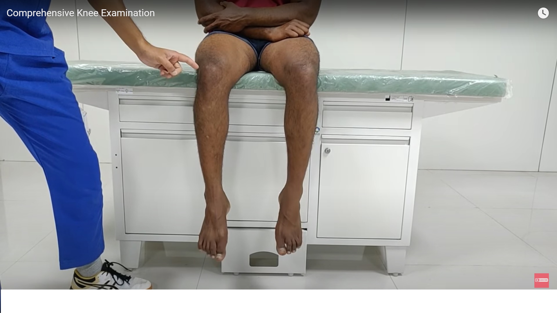
Clinical Examination
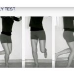
Gait Assessment
-
No obvious varus thrust during walking
-
Ligaments appeared clinically stable during gait analysis
Alignment
-
Visible varus deformity present during standing examination
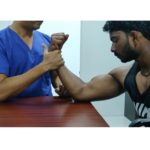
Ligament Stability
-
Varus and valgus stress testing performed in:
-
Full extension
-
Ten to twenty degrees of knee flexion
-
Key Observation
-
The varus deformity could be partially corrected manually during flexion.
This suggests that:
-
Some component of the deformity may be intra-articular, often due to medial joint space narrowing and meniscal extrusion.
Rotational Assessment
The patient was also evaluated in the prone position to assess:
-
Hip rotational profile
-
Tibial torsion
-
Femoral rotational alignment
Findings included:
-
Approximately forty degrees of hip internal and external rotation
-
Foot progression angle around thirty degrees
-
No abnormal tibial or femoral torsion
This confirmed that the primary deformity was likely in the coronal plane rather than rotational.
Radiographic Evaluation
Standard Radiographs
Radiographic evaluation included:
-
Patellar skyline view
-
Lateral knee radiograph
-
Long-leg alignment radiograph
Key observations:
-
Patella appeared well centered
-
No major degenerative changes visible on lateral radiograph
Mechanical Axis Assessment
The mechanical axis line was drawn connecting:
-
Center of the femoral head
-
Center of the ankle joint
This line determines the location of the load-bearing axis.
Interpretation
-
The mechanical axis passed medial to the knee center
-
This confirmed varus alignment.
Deformity Analysis
To determine the origin of deformity, two key angles were measured:
Mechanical Lateral Distal Femoral Angle
-
Measured between:
-
Femoral mechanical axis
-
Distal femoral joint line
-
Medial Proximal Tibial Angle
-
Measured between:
-
Tibial mechanical axis
-
Proximal tibial joint line
-
Normal Reference Value
-
Approximately eighty-seven degrees for each angle.
Findings
Measured angles revealed:
-
Femoral angle around ninety-three degrees
-
Tibial angle around eighty-one degrees
Interpretation:
-
Both femur and tibia contributed to the varus deformity.
Surgical Planning
Because deformity existed in both bones, a double-level osteotomy was planned.
Planned Corrections
-
Closing wedge distal femoral osteotomy
-
Opening wedge proximal tibial osteotomy
The goal was to shift the mechanical axis to approximately fifty-two to fifty-three percent of the tibial plateau width.
Importance of Joint Line Orientation
Correcting only one bone in a double-level deformity may lead to:
-
Excessive joint line obliquity
-
Abnormal joint biomechanics
Maintaining physiological joint line orientation is critical for:
-
Joint stability
-
Long-term cartilage health
Leg Length Considerations
Opening wedge osteotomy can increase limb length due to:
-
Straightening of the limb
-
Opening of the osteotomy gap
Closing wedge osteotomy may shorten the limb slightly.
Balancing both procedures can help maintain appropriate leg length.
Indications for Double-Level Osteotomy
A useful rule:
-
When the mechanical axis does not intersect the tibial plateau, a double-level osteotomy should be considered.
Role of Arthroscopy During Osteotomy
Routine arthroscopy before osteotomy remains debated.
Possible indications include:
-
Mechanical symptoms
-
Loose bodies
-
Symptomatic cartilage flaps
Many surgeons now perform selective arthroscopy rather than routine arthroscopy.
Case Two
Osteochondral Defect of the Lateral Femoral Condyle
Patient Profile
-
Twenty-six-year-old professional wrestler
-
Persistent knee pain following previous cartilage surgery
Previous Treatment
-
Arthroscopy with microfracture procedure
Current Symptoms
-
Pain during walking and squatting
-
Recurrent swelling
-
Occasional locking
-
Inability to return to sport
Clinical Examination
Key Findings
-
Mild quadriceps muscle wasting
-
Pain during deep knee flexion
-
Sharp tenderness over the lateral femoral condyle
Functional Testing
-
Pain reproduced during mini squat around twenty to thirty degrees of flexion.
Imaging Findings
Magnetic resonance imaging demonstrated:
-
Failed microfracture repair
-
Large osteochondral defect of the lateral femoral condyle
-
Subchondral cyst formation
Estimated lesion size:
-
Approximately twenty by twelve millimetres
This confirmed involvement of the osteochondral unit rather than cartilage alone.
Surgical Strategy
Because the subchondral bone was involved, treatment required osteochondral reconstruction.
Preferred Option
-
Osteochondral autograft transfer procedure
Multiple graft plugs may be required due to lesion size.
Additional Biological Augmentation
Remaining defect areas may be filled with:
-
Cartilage matrix scaffold
-
Collagen membrane
-
Fibrin glue
These techniques aim to enhance biological repair.
Case Three
Failed Anterior Cruciate Ligament Reconstruction
Patient Profile
-
Twenty-four-year-old male
-
Recreational badminton player
Injury History
-
Initial anterior cruciate ligament reconstruction
-
Reinjury three months before presentation
Current Symptoms
-
Persistent instability
-
Difficulty with pivoting movements
-
Lack of confidence during activities
Clinical Examination
Range of Motion
-
Mild loss of extension due to locked medial meniscus tear
Stability Tests
Positive findings included:
-
Grade three Lachman test
-
Positive anterior drawer test
-
Positive pivot shift test
Posterolateral Corner Testing
Testing demonstrated mild laxity but not significant instability.
Imaging Evaluation
Radiographic Alignment
-
Coronal alignment appeared normal
Magnetic Resonance Imaging
Findings included:
-
Absent anterior cruciate ligament graft
-
Bucket-handle tear of the medial meniscus
Importance of Posterior Tibial Slope
Posterior tibial slope influences anterior tibial translation.
Increased slope can significantly increase:
-
Stress on anterior cruciate ligament grafts
This increases the risk of graft failure.
Computed Tomography Analysis
Computed tomography was performed to assess:
-
Tunnel size
-
Tunnel position
-
Implant placement
Findings included:
-
Tibial tunnel approximately twelve millimetres in diameter
-
Femoral tunnel approximately fourteen millimetres
Tunnel positions were considered acceptable for revision surgery.
Revision Surgery Planning
Key surgical decisions included:
Graft Choice
Autograft was preferred for revision reconstruction.
Tunnel Management
-
Single-stage revision considered feasible
-
Tunnel enlargement did not mandate staged reconstruction
Additional Procedures
Planned procedures included:
-
Anterior closing wedge osteotomy to reduce tibial slope
-
Lateral extra-articular tenodesis for rotational stability
Meniscus Management
The patient also had a bucket-handle medial meniscus tear.
Intraoperative decision-making would determine whether to:
-
Repair the meniscus
-
Perform partial meniscectomy
Loss of medial meniscus function may increase instability risk.
Key Lessons from These Cases
-
Accurate deformity analysis is essential before performing osteotomy.
-
Double-level osteotomy helps maintain normal joint line orientation.
-
Failed cartilage procedures require treatment of the entire osteochondral unit.
-
Posterior tibial slope plays an important role in anterior cruciate ligament stability.
-
Revision anterior cruciate ligament reconstruction often benefits from additional stabilizing procedures.

Leave a Reply