Courtesy: Dr. Anant Joshi, Dr Ashok Shyam, Ortho TV
Hidden and Missed Lesions in Knee Arthroscopy
Introduction
Knee arthroscopy is one of the most commonly performed orthopedic procedures. Despite advances in arthroscopic techniques and imaging, important lesions may still be missed during surgery. These missed lesions can lead to persistent symptoms, failed reconstructions, recurrent instability, and long-term joint degeneration.
Lesions may be missed because of:
- Truly hidden pathology
- Inexperience of the surgeon
- Inadequate visualization
- Failure to systematically examine all compartments
- Overconfidence leading to incomplete assessment
Clinical Importance of Missing Lesions
Missed pathology can significantly affect both short-term and long-term outcomes.
Immediate Consequences
Persistent Symptoms
Examples include:
- Locking due to missed loose bodies
- Ongoing pain from untreated cartilage lesions
Short-Term Consequences
Progression of Meniscal Tears
A small missed meniscal tear may later progress into:
- Bucket-handle tear
- Mechanical symptoms
- Irreparable meniscal damage
Long-Term Consequences
Persistent Instability
Missed collateral or posterior stabilizer injuries may lead to:
- Failure of ACL reconstruction
- Residual instability
- Degenerative arthritis
Serious Diagnostic Errors
One major pitfall is performing ACL reconstruction in a PCL-deficient knee.
This occurs when a PCL injury is missed and the lax ACL appearance is incorrectly interpreted as ACL insufficiency.
Truly Hidden Lesions
Loose Bodies
Loose bodies can be difficult to identify because they may hide within:
- Synovial folds
- Gutters
- Popliteal recesses
Some loose bodies may require a posterolateral portal for retrieval.
Intrasubstance Meniscal Tears
These tears may not be visible on the meniscal surface initially.
Diagnostic Features
- Meniscus may appear normal externally
- Tear becomes evident only after trimming or probing
Common Associations
- Horizontal cleavage tears
- Parameniscal cysts
Parameniscal cysts develop due to extrusion of synovial fluid through the tear.
Lesions Missed Due to Inexperience or Technical Errors
Posterior Cruciate Ligament (PCL) Injuries
PCL injuries are frequently missed because the ligament may appear covered by intact synovium.
Important Principle
The synovium should be carefully shaved to properly visualize the PCL.
The “Sloppy ACL” Sign
In PCL deficiency:
- The ACL may appear lax or redundant
- This can be mistaken for ACL insufficiency
This diagnostic error may lead to inappropriate ACL reconstruction instead of treating the actual PCL injury.
Importance of Probing
The probe is one of the most important diagnostic instruments during arthroscopy.
Uses of Probing
Probing helps detect:
- Hidden meniscal tears
- Meniscal instability
- Cartilage softening
- Flap tears
- Subtle instability
Failure to adequately probe structures is a common reason lesions are missed.
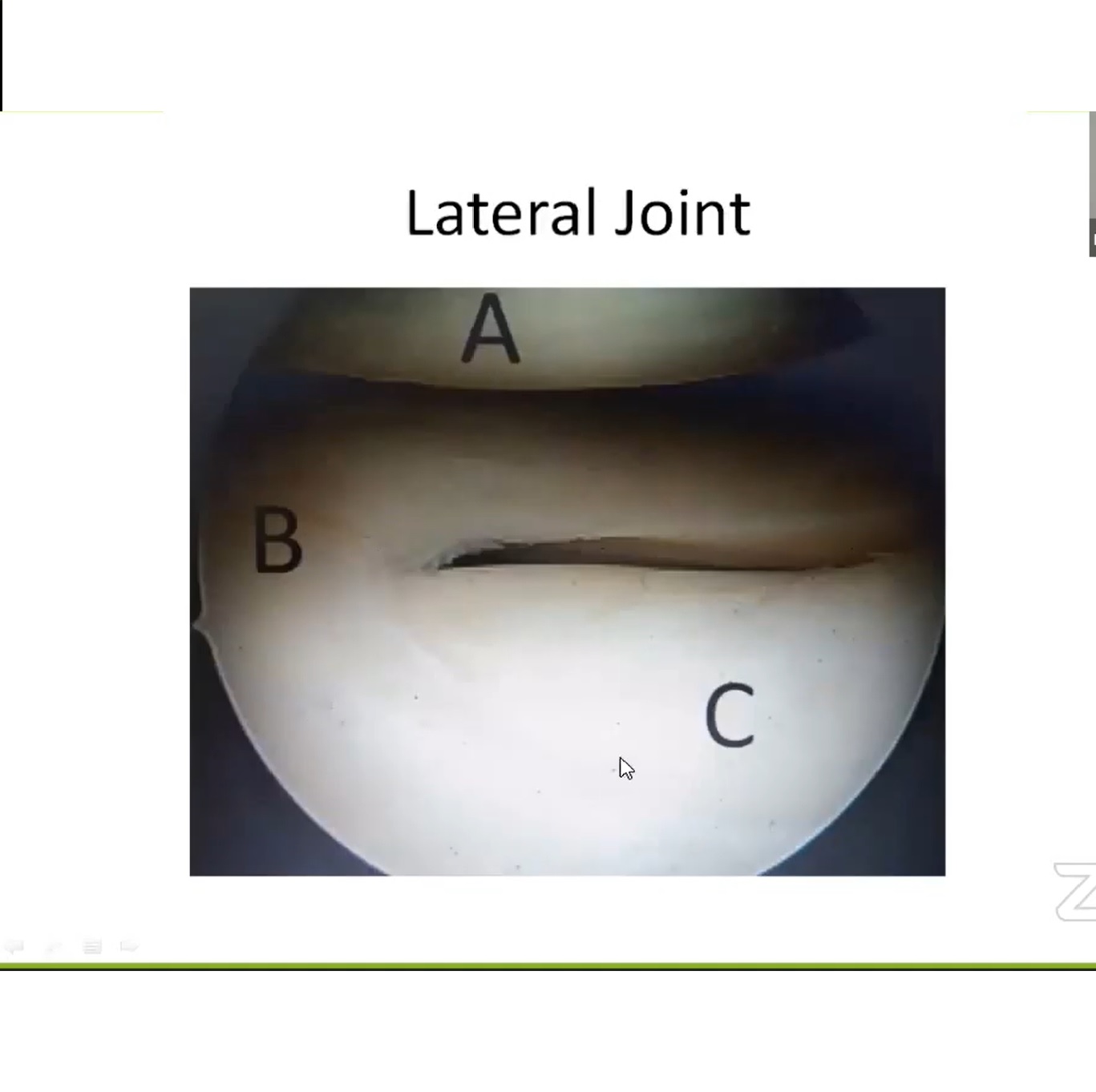
Articular Cartilage Lesions
MRI Limitations
MRI may fail to detect:
- Cartilage softening
- Early chondral degeneration
Arthroscopic Diagnosis
Cartilage lesions are often diagnosed by:
- Direct visualization
- Arthroscopic palpation with probe
A cartilage surface may appear normal visually but feel soft on probing and represent the true source of pain.
Missed Meniscal Tears
Tears Missed Without Probing
Some meniscal tears become apparent only after probing reveals:
- Displacement
- Instability
- Hidden cleavage
Hidden Flap Tears
A meniscal flap may fold beneath the meniscus and remain concealed.
Diagnosis
Careful probing is required to identify the displaced fragment.
Missed ACL Lesions
Femoral-Side ACL Tears
Certain ACL tears may be missed unless:
- The knee is placed in the figure-of-four position
- The femoral attachment is carefully inspected
Without appropriate positioning, the ACL may appear intact.
Importance of Joint Positioning
Proper arthroscopic evaluation requires continuous adjustment of both:
- Scope position
- Knee position
Some lesions become visible only in specific positions.
Example
Posterior femoral condyle cartilage lesions may only be visualized when the knee is flexed.
Posterior Compartment: The Most Commonly Missed Area
Many surgeons fail to routinely inspect the posterior compartment.
This area commonly contains important hidden lesions including:
- Meniscal root tears
- Ramp lesions
- Posterior capsular injuries
Ramp Lesions
Definition
Ramp lesions are tears between:
- Posterior horn of the medial meniscus
- Posteromedial capsule
Association with ACL Injuries
Ramp lesions commonly occur with ACL tears and may contribute to persistent instability.
Types
Visible Ramp Lesions
Seen directly during standard arthroscopy.
Hidden Ramp Lesions
Require probing through the posteromedial capsule for diagnosis.
Diagnostic Technique
The arthroscope is advanced between:
- Posterior cruciate ligament
- Medial femoral condyle
This allows inspection of the posteromedial compartment.
Clinical Importance
If missed, ramp lesions may lead to:
- Persistent instability
- ACL graft failure
- Recurrent pivot shift
Meniscal Root Tears
Types of Root Tears
Medial Root Tears
- Usually degenerative
- Common in middle-aged patients
Lateral Root Tears
- Usually traumatic
- Frequently associated with ACL injuries
Biomechanical Importance
A root tear effectively renders the meniscus nonfunctional.
Consequences include:
- Meniscal extrusion
- Increased joint contact pressure
- Accelerated degeneration
Biomechanically, it behaves similarly to a total meniscectomy.
Treatment
Healthy meniscal root tears should generally be repaired.
Common Technique
- Sutures passed through root
- Fixation via transtibial tunnel
Popliteus Tendon Lesions
Importance
Popliteus tendon injuries are frequently underdiagnosed.
Arthroscopic Findings
Possible findings include:
- Hemorrhagic tendon
- Synovial sheath tear
- Femoral avulsion
Treatment
Management options include:
- Arthroscopic repair
- Open repair
- Popliteus sling reconstruction in severe cases
Hidden Synovial Lesions
Certain lesions may remain concealed beneath synovium.
Examples
- Ganglion cysts
- Pigmented villonodular synovitis (PVNS)
- Synoviomas
Detection
These lesions may require synovial shaving for adequate visualization.
Key Surgical Principles
To avoid missing important pathology, surgeons should:
- Systematically inspect all compartments
- Use the probe extensively
- Dynamically reposition the knee
- Routinely examine the posterior compartment
- Carefully inspect meniscal roots and ramp areas
Conclusion
Hidden lesions in knee arthroscopy are common and clinically significant. Failure to recognize these lesions may result in:
- Persistent pain
- Mechanical symptoms
- Recurrent instability
- ACL graft failure
- Early osteoarthritis
A systematic arthroscopic approach, careful probing, dynamic joint positioning, and thorough posterior compartment evaluation are essential for improving patient outcomes and preventing long-term complications.
Related Posts
 Portals in Knee arthroscopy
Courtesy: Dr David Rajan, Arthroscopy Course, Coimbatore
 Diagnostic Knee arthroscopy and ACL Reconstruction
Courtesy: Saseendar Sundaram, Neeraj Srivastava
 MCQ Sports Knee
Answer: https://bit.ly/orthomcq3 (Copy and Paste in Browser)
Leave a Reply