• Skip to main content
  • Skip to secondary menu
  • Skip to primary sidebar
OrthopaedicPrinciples.com

OrthopaedicPrinciples.com

Integrating Principles and Evidence

Integrating Principles and Evidence

  • Home
  • Editorial Board
  • Our Books
    • Evidence Based Orthopaedic Principles
  • Courses
  • Exams
  • Reviews
  • Live Program
  • Contact

IntraArticular Fractures of the Calcaneus

Courtesy: Prof Dane Wukich MD, CHarles Gregory Distinguished Chair in Orthopaedic Surgery, UT SouthWestern, Dallas, Texas

Historical perspective
• Norris (1839) and Malgaigne (1843) described calcaneal fractures
• Lorenz Bohler (1935) described the mechanism of injury and classified them

Epidemiology
• 50-60% of all tarsal bone fractures
• <10% are open injuries
• Mainly in young adults (20-39 years of age)

Mechanism of injury
• High energy injury usually by fall from height or motor vehicle accidents
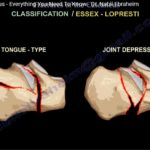
Classification

Essex- Lopresti classification

  • x-ray based classification system
  • 2 main types- joint depression type & tongue type

Sander’s classification

• CT scan based classification
• Most widely used and has prognostic value
• Guide the treatment

Look at the level of widest portion of posterior facet of talus in coronal section

Type 1 – undisplaced (non-surgical management is preferred)
Type 2 – two fragments (surgical fixation)
Type 3 – three fragments (surgical fixation)
Type 4 – four fragments (consider primary fusion with reduction)
A – lateral 1/3 rd.
B – middle 2/3 rd.
C – medial 1/3 rd.

Initial treatment

• Pain relief
• Control oedema (ice, elevation, compression and splinting)
• Urgent irrigation and debridement along with wound management in open injuries
Important radiographic parameters

  • Bohler’s angle- normally 200 to 400
  • Shenton’s line- normal gradual curve on the medial side
  • The constant fragment – helps in restoring the anatomy

Note: look for wrinkle sign before proceeding to surgery
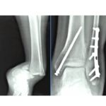
Goals of surgery

• Regain length
• Correct varus
• Reduce the joint
• Restore height

Lateral extensile approach

• Primary approach for many years
• Allows adequate visualization
• Allows manipulation to restore alignment
• Creating full thickness flap

Risk factors for wound complications
– Diabetes
– Smoking
– Open fractures

Minimally invasive techniques

• Sinus tarsi approach
• Percutaneous techniques
• External fixation

Sinus tarsi approach
– Increasing in popularity
– Lesser wound complications
– Technically challenging
– Lateral position
– Half pin used as a joystick

Conclusion

• Calcaneal fracture is a life altering event
• Goal is to improve function and avoid complications
• Know which patient needs surgery
• Secondary reconstruction to correct malunions are highly challenging


Post Views: 8,844

Related Posts

  • Difficult Ankle Fractures

    Courtesy: Saqib Rehman MD, Director of Orthopaedic Trauma, Temple University, Philadelphia, Pennsylvania, USA

  • Calcaneal Fractures Part 1

  • Fractures of the Calcaneus

    Courtesy: Prof Nabil Ebraheim, University of Toledo, Ohio, USA It could be open or closed.…

Reader Interactions

Leave a Reply

Your email address will not be published. Required fields are marked *

Primary Sidebar

Follow Us

instagram slideshare

Categories

  • -Applied Anatomy
  • -Approaches
  • -Basic Sciences
  • -Cartilage & Meniscus
  • -Classifications
  • -Examination
  • -Foot and Ankle
  • -Foot and Ankle Trauma
  • -FRCS(Tr and Orth) tutorials
  • -Gait
  • -Hand and Wrist
  • -Hand and Wrist Trauma
  • -Hand Infections
  • -Hip and Knee
  • -Hip Preservation
  • -Infections
  • -Joint Reconstruction
  • -Knee Arthroplasty
  • -Knee Preservation
  • -Metabolic Disorders
  • -Oncology
  • -OrthoBiologics
  • -OrthoPlastic
  • -Paediatric Orthopaedics
  • -Paediatric Trauma
  • -Patellofemoral Joint
  • -Pelvis
  • -Peripheral Nerves
  • -Principles
  • -Principles of Surgery
  • -Radiology
  • -Rheumatology
  • -Shoulder and Elbow
  • -Shoulder and Elbow Arthroplasty
  • -Spine Deformity
  • -Spine Oncology
  • -Spine Trauma
  • -Spine, Pelvis & Neurology
  • -Sports Ankle and Foot
  • -Sports Elbow
  • -Sports Knee
  • -Sports Medicine
  • -Sports Medicine Hip
  • -Sports Shoulder
  • -Sports Wrist
  • -Statistics
  • -Technical Tip
  • -Technology in Orth
  • -Trauma
  • -Trauma (Upper Limb)
  • -Trauma Life Support
  • -Trauma Reconstruction
  • Book Shelf
  • Book Shelf Medical
  • Careers
  • Case Studies and Free Papers
  • DNB Ortho
  • Evidence Based Orthopaedic Principles
  • Evidence Based Orthopaedics
  • Exam Corner
  • Fellowships
  • Guest Editor
  • Guest Reviews
  • Image Quiz
  • Instructional Course Lectures
  • Journal Club
  • MCQs
  • Meetings and Courses
  • Multimedia
  • News and Blog
  • Plaster Techniques
  • Podcasts
  • Public Health
  • Rehabilitation
  • Research
  • Shorts and Reels
Copyright@orthopaedicprinciples.com. All right rerserved.