Infection of the finger is a common problem that can vary in severity. Serious infection of the fingers may require urgent surgical care.
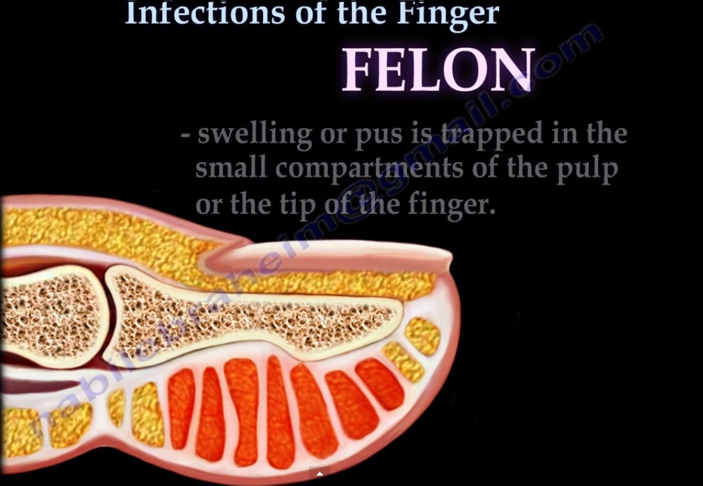
Felon
•Deep infection of the soft pad, or pulp of the fingertip. Usually the result of a puncture wound.
•Swelling or pus is trapped in the small compartments of the pulp or the tip of the finger.
Symptoms:
•Unusual redness or swelling
•Throbbing pain at the tip of the finger
•Firm swelling
•Visible pus
If the infection goes untreated, it may lead to severe symptoms such as skin necrosis, flexor tenosynovitis, osteomyelitis and arthritis of the distal interphalangeal joint.
Treatment:
•antibiotics if the infection is caught early.
•Surgery is the usual treatment: incision and drainage of the felon.
Paronychia
•Infection involving the soft tissues around the fingernail. Most common bacterial infection of the hand and is often associated with a simple hangnail.
Symptoms:
•Swelling
•Redness
•Pus formation
•Pain in the soft tissue around the nail plate.
Treatment:
•antibiotics if the infection is caught early.•Surgery is the usual treatment: incision and drainage with or without partial nail removal for subungal abcess.
Herpetic whitlow
•Painful infection caused by the herpes simplex virus that usually affects the fingers or thumb.
•Commonly contracted by dental workers and medical workers exposed to oral secretions and can occur in infants.
Symptoms
•Swelling and tenderness
•Redness
•Vesicle formation
•Fever
•Swollen lymph nodes
Treatment
•Conservative: the infection is self-limiting. Antiviral treatments applied to the skin (Acyclovir). Antibiotics are not used unless secondary infection is present. Drainage of the vesicles may lead to viral encephalitis.
Flexor tenosynovitis
•Relatively common infection of the hand usually caused by staphylococcus aureus. Usually occurs due to prior penetrating trauma and infection. The index, middle, and ring fingers are most commonly affected.
Symptoms
•Painful swelling of the finger that hurts worse with motion.
Kanavel’s four cardinal signs:
1-Uniform swelling of the entire finger
2-The finger is flexed
3-Intense pain when attempting to straighten the finger. occurs early.
4-Tenderness along the course of the tendon sheta. Most important sign.
Treatment
•If infection is caught early: IV antibiotics
•If infection is severe: open drainage. Early drainage of the infection to avoid skin loss, tendon necrosis, and osteomyelitis. The posterolateral incision is better than a zig-zag incision. Avoid indwelling catheter.
Infection may spread from the tendon into the deep palmar space or to the Parona’s space in the forearm. The little finger communicates with the ulnar bursa. The thumb communicates with the radial bursa. The radial and ulnar bursa communicate proximal to the carpal tunnel. Infection may travel from the little finger into the ulnar bursa to the parona’s space. Infection can also travel from the thumb into the radial bursa to the parona’s space. Infection may cause “horse shoe” tenosynovitis. Infection travels from the thumb through the radial bursa to the ulnar bursa infecting the little finger. may need combination of incisions for drainage


i like it
beautiful presentation