
Courtesy: Dr T Vail, Ashok Shyam TV, Ortho
Imaging of the Hip
1. Why Hip Imaging Matters
Hip imaging is not simply about identifying pathology. It helps us understand:
- Hip morphology
- Hip biomechanics
- Relationship between the femur and pelvis
- Leg length
- Femoral offset
- Acetabular coverage
- Cartilage and labral pathology
- Femoroacetabular impingement
- Wear and osteolysis
The imaging findings should therefore be considered in the context of hip reconstruction and biomechanics.
2. Plain Radiography
Plain radiographs provide important information about hip morphology and alignment.
Important parameters include:
- Acetabular coverage
- Femoral head position
- Lateral and superior subluxation
- Leg length
- Femoral neck-shaft angle
- Femoral offset
- Acetabular morphology
These measurements are particularly useful when planning hip reconstruction or arthroplasty.
3. Important Radiographic Measurements
Acetabular Coverage
Several radiographic indices are used to assess how much of the femoral head is covered by the acetabulum.
Index of Elevation
Measured from a horizontal reference line to the lateral edge of the acetabulum.
Extrusion Index
Represents the proportion of the femoral head that is uncovered by the acetabulum.
The relationship between the covered and uncovered portions of the femoral head can therefore be assessed on an AP radiograph.
4. Teardrop
The teardrop is an important and relatively reliable landmark on the AP pelvis.
It is useful because it accounts relatively well for:
- Pelvic rotation
- Pelvic tilt
Measurements based on the teardrop can therefore be useful when comparing radiographs over time.
It can help assess:
- Lateral subluxation
- Superior subluxation
- Hip reconstruction
5. Femoral Morphology
Important measurements include:
Femoral Neck-Shaft Angle
Helps define femoral morphology and is relevant when assessing reconstruction.
Femoral Offset
An important biomechanical parameter.
It can be relevant to:
- Impingement
- Component positioning
- Hip resurfacing
The lecture mentions that femoral offset can be measured on a frog-leg view.
6. Shenton’s Line
Shenton’s line is commonly used to assess hip alignment, particularly in developmental dysplasia of the hip.
It helps determine whether the femoral head is anatomically aligned with the acetabulum or displaced.
7. Crossover Sign
The crossover sign is an important radiographic indicator related to acetabular morphology and possible impingement.
On an AP pelvis:
- The anterior acetabular rim is traced.
- The posterior acetabular rim is traced.
- Crossover of the anterior rim over the posterior rim suggests potential anterior impingement.
Important point
The crossover sign is influenced by:
- Pelvic position
- Pelvic rotation
- Lumbar lordosis
Therefore, it should be interpreted in the appropriate clinical and radiographic context.
8. Cartilage and Labral Pathology
Plain radiographs have important limitations.
They are poor at identifying:
- Isolated cartilage defects
- Cartilage delamination
- Subtle chondral injuries
- Many loose bodies
The lecture emphasizes that plain radiographs detect only a proportion of loose bodies.
This is important because the amount of cartilage damage and degeneration has major implications for the success of hip-preserving procedures.
9. MRI
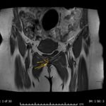
MRI provides substantially more information than plain radiography for soft-tissue and cartilage pathology.
It allows assessment of:
- Labrum
- Articular cartilage
- Bone
- Soft tissues
- Chondral defects
- Cartilage delamination
MRI is therefore particularly useful when the clinical question involves labral or chondral pathology.
10. MR Arthrography
MRI with intra-articular gadolinium contrast improves the ability to diagnose labral pathology.
It provides better visualization of:
- Labral tears
- Cartilage surfaces
- Intra-articular pathology
The lecture particularly emphasizes the improved diagnostic accuracy of contrast-enhanced MRI for labral pathology.
11. Imaging in Perthes Disease
Plain radiographs may demonstrate the obvious bony abnormalities of Perthes disease, but they may not adequately demonstrate:
- Cartilage delamination
- Chondral damage
- Absence of viable articular cartilage
These findings can be important in determining whether a hip-preserving procedure is likely to succeed.
12. Imaging and Hip Arthroscopy
The presence of:
- Degeneration
- Osteonecrosis
- Cartilage damage
is associated with poorer outcomes following hip arthroscopy.
Therefore, imaging is important not only for diagnosis but also for patient selection and prognosis.
13. Femoroacetabular Impingement
Imaging should identify not only the labral tear but also the underlying morphology responsible for the pathology.
This is an important principle:
Treating the labral tear alone may not be sufficient if the underlying impingement morphology remains uncorrected.
Hips with labral tears are frequently associated with underlying morphological abnormalities.
14. Cam Lesions
Newer MRI techniques, including radial imaging, can better demonstrate the shape of the femoral head-neck junction.
They can identify:
Cam lesions
These appear as a prominence or bump at the:
- Femoral head-neck junction
- Adjacent metaphyseal region
This morphology can contribute to:
- Femoroacetabular impingement
- Labral pathology
- Chondral delamination
Therefore, identifying the morphology is important for appropriate treatment.
15. Imaging Modality: Practical Comparison
| Imaging | Main Information |
|---|---|
| Plain radiograph | Alignment, morphology, coverage, offset, subluxation |
| AP pelvis | Acetabular morphology, Shenton’s line, crossover sign |
| Frog-leg view | Femoral morphology and offset |
| MRI | Cartilage, labrum, bone and soft tissues |
| MR arthrography | Improved assessment of labral/intra-articular pathology |
| Radial MRI | Femoral head-neck morphology and cam lesions |
Key Clinical Pearls
- Hip imaging should be interpreted in terms of biomechanics and reconstruction, not simply pathology.
- Teardrop is an important reliable landmark on AP radiographs.
- Shenton’s line helps assess hip alignment, particularly in DDH.
- Crossover sign suggests acetabular morphology associated with potential anterior impingement.
- Femoral offset is important for biomechanics and reconstruction.
- Plain radiographs are poor for detecting isolated cartilage defects and subtle chondral pathology.
- MRI provides much better assessment of cartilage, labrum, bone and soft tissues.
- MR arthrography improves detection of labral pathology.
- Cam morphology can be better assessed with radial MRI.
- In a patient with a labral tear, look for the underlying morphology causing the tear, particularly impingement.
- The degree of cartilage degeneration has important prognostic implications for hip-preserving surgery.


Leave a Reply