Courtesy: Prof Nabil Ebraheim, University of Toledo, Ohio, USA
Introduction
-
Heel pain is an extremely common clinical complaint.
-
It has several distinct and overlapping causes.
-
Accurate identification of the underlying cause is essential to ensure appropriate and effective treatment.
-
Overlapping pain locations often make diagnosis challenging and, at times, confusing.
Common Causes of Heel Pain
The commonly encountered causes of heel pain include:
-
Plantar fasciitis
-
Baxter’s nerve compression
-
Fat pad atrophy
-
Achilles tendinitis
-
Haglund’s deformity
-
Stress fracture of the calcaneus
-
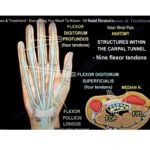
Tarsal tunnel syndrome
-
Lumbosacral spine radiculopathy
Clinical Causes and Their Characteristics
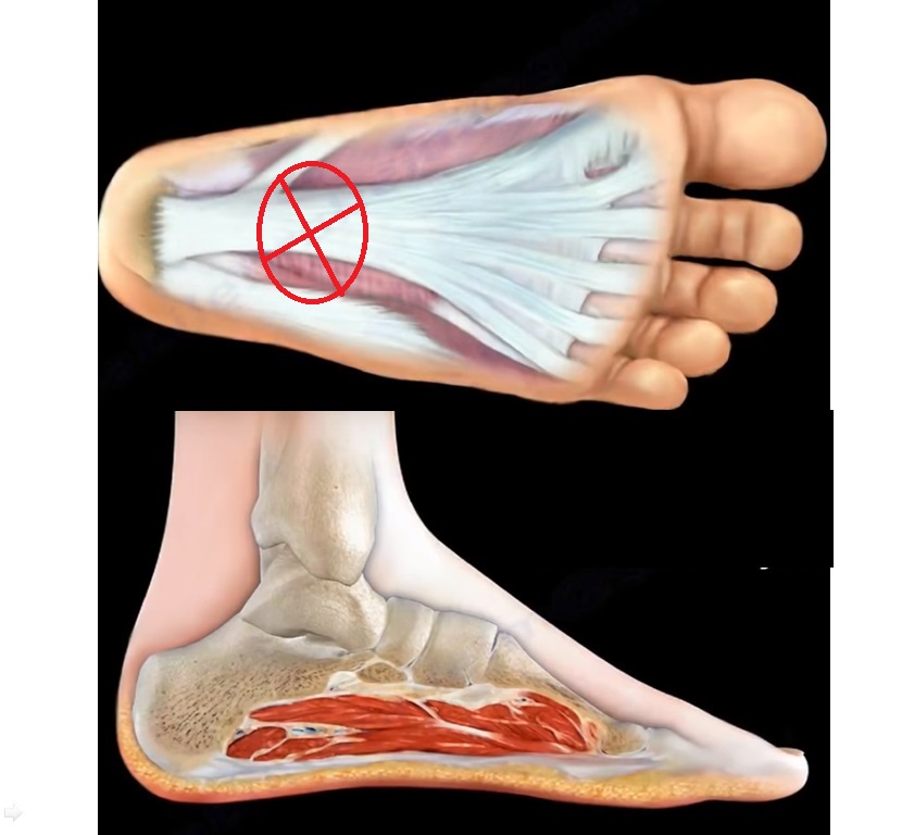
1. Plantar Fasciitis
-
Caused by irritation and inflammation of the thick fibrous tissue on the plantar aspect of the foot.
-
Pain is most severe during the first steps taken in the morning, commonly referred to as start-up pain.
-
Pain persists with activity throughout the day and intensifies with prolonged standing or exercise.
-
Examination findings include:
-
Tenderness over the plantar medial aspect of the heel
-
Medial calcaneal tenderness
-
Negative Tinel’s sign
-
-
Radiographs may demonstrate a plantar heel spur.
-
Frequently associated with a tight Achilles tendon.
Treatment options include:
-
Night splints
-
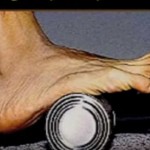
Physical therapy
-
Cushioned silicone heel inserts
-
Corticosteroid therapy
-
Achilles tendon stretching exercises
2. Baxter’s Nerve Compression
-
Baxter’s nerve is the first branch of the lateral plantar nerve.
-
It accounts for approximately 20 percent of heel pain cases.
-
This nerve provides motor innervation to the abductor digiti minimi muscle.
-
Compression of this nerve often produces symptoms similar to plantar fasciitis.
-
The nerve travels between the abductor hallucis and quadratus plantae muscles, then turns sharply laterally beneath the calcaneus.
-
Pain is typically localized to the medial plantar aspect of the heel.
-
This condition is frequently overlooked or misdiagnosed, particularly in athletes.
3. Fat Pad Atrophy
-
The heel fat pad normally cushions the calcaneus during weight bearing.
-
In fat pad atrophy, thinning of this tissue leads to loss of cushioning and increased pain.
-
Commonly seen in elderly patients.
-
There is often a history of repeated corticosteroid injections for plantar fasciitis.
-
Pain characteristics include:
-
Deep, central plantar heel pain
-
Non-radiating pain
-
Worsening when barefoot
-
Improvement when walking on toes
-
-
Examination reveals tenderness at the central aspect of the heel pad.
4. Achilles Tendinitis
-
Typically presents as a chronic condition with symptoms lasting several months.
-
Patients report pain and swelling at the posterior aspect of the ankle.
-
Physical examination reveals:
-
Thickening of the Achilles tendon
-
Tenderness near its insertion on the calcaneus
-
5. Haglund’s Deformity
-
Characterized by insertional calcification and bony prominence at the Achilles tendon insertion.
-
Initial management includes:
-
Physical therapy
-
Anti-inflammatory medication
-
-
Injections should be administered around the tendon and not through the tendon substance.
-
If symptoms persist beyond 6 months, surgical intervention may be required.
-
Surgical procedures may include:
-
Excision of the posterosuperior calcaneal prominence
-
Removal of insertional calcification
-
-
If more than 50 percent of the Achilles tendon is detached during surgery:
-
Tendon fixation to the calcaneus is required using suture anchors
-
Tendon transfer may be necessary
-
-
Advanced degeneration may require Achilles tendon debridement, calcaneal exostectomy, and flexor hallucis longus tendon transfer.
6. Stress Fracture of the Calcaneus
-
Occurs due to repetitive overuse.
-
Patients experience severe weight-bearing heel pain that worsens with walking and running.
-
Pain does not improve during the day and is present with each step.
-
Medial to lateral compression of the calcaneal tuberosity reproduces pain.
-
Radiographs may appear normal in early stages.
-
Magnetic resonance imaging is often required for definitive diagnosis.
7. Tarsal Tunnel Syndrome
-
Characterized by numbness and paresthesia in the plantar aspect of the foot.
-
Symptoms worsen with activity and may awaken the patient at night.
-
A ganglion cyst is a known cause of compression within the tarsal tunnel.
-
Magnetic resonance imaging should be evaluated to identify space-occupying lesions.
-
Surgical excision of the ganglion often leads to favorable outcomes and symptom resolution.
8. Lumbosacral Spine Radiculopathy
-
Lateral foot pain may originate from lumbosacral disc pathology.
-
Herniation at the L5–S1 level commonly affects the S1 nerve root.
-
S1 nerve root involvement results in:
-
Pain along the lateral aspect of the foot
-
Decreased sensation in the same distribution
-
Leave a Reply