
Courtesy: Sreenadh Gella, FRCSOrth, Birmingham, UK
Hand Infections and Spaces of the Hand
1. Basic Concept
The spaces of the hand are potential spaces that become clinically apparent when they contain:
- Infection
- Pathological fluid collection
- Tumour
- Reactive effusion
Infections commonly begin as cellulitis. They may resolve spontaneously or progress to an abscess or deeper collection.
Deep hand infections are particularly important in patients with:
- Diabetes
- Immunosuppression
- Other significant medical comorbidities
The most common organism mentioned is Staphylococcus aureus. Animal bites are commonly polymicrobial.
2. Important Hand Spaces
Superficial spaces
- Pulp spaces
- Web spaces
- Dorsal subcutaneous space
- Dorsal subaponeurotic space
Deep spaces
- Flexor tendon sheaths
- Radial bursa
- Ulnar bursa
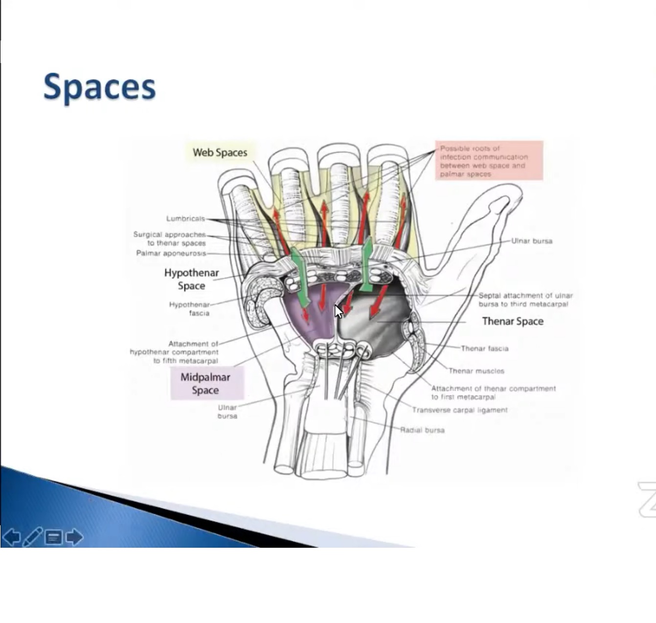
- Mid-palmar space
- Thenar space
- Hypothenar space
- Space of Parona
Deep infections may take longer to become clinically obvious.
3. Flexor Sheath Infection
Flexor sheath infection is commonly caused by:
- Penetrating injury
- Direct inoculation
- Continuous spread from surrounding tissues
Indirect spread is less common.
An important anatomical variation is communication between the little finger flexor sheath and the ulnar bursa. Communication between the radial and ulnar bursae may also occur.
Clinical Features
The classic presentation includes:
- Sausage-shaped swelling
- Finger held in flexion
- Tenderness along the flexor sheath
- Pain on passive stretching
These are the classic Kanavel signs.
A positive combination of these findings should make you highly suspicious of flexor sheath infection.
4. Treatment of Flexor Sheath Infection
Early infection
Nonoperative treatment with:
- IV antibiotics
- Close observation
may be considered in selected early cases.
However, the preferred approach when there is significant suspicion is generally:
Incision and drainage with washout
This is intended to reduce the risk of:
- Severe stiffness
- Persistent infection
- Further complications
Continuous irrigation may also be used in some units.
Complications
Complication rates may reach 30 to 40% and include:
- Stiffness
- Recurrent infection
- Further surgery
- Scar problems
5. Poor Prognostic Factors
Important factors suggesting a poorer outcome include:
- Diabetes
- Human or animal bite
- Subcutaneous purulence
- Digital ischaemia
- Delayed presentation
- Severe infection
These factors should make you more aggressive in your assessment and management.
6. Late Complications
Even after the infection has settled, significant problems may remain.
Stiffness
Initial treatment is:
- Hand therapy
- Passive range of movement
- Active range of movement
The lecture recommends prolonged therapy, with surgery considered if stiffness persists.
Tendon necrosis and rupture
Infection can cause vascular compromise and tendon necrosis, resulting in:
- Late tendon rupture
- Severe functional impairment
These cases may require two-stage reconstruction, although outcomes can be poor.
7. Space of Parona Infection
Spread of infection from the flexor sheath into the:
- Ulnar bursa
- Radial bursa
- Space of Parona
is an important complication.
This is considered a surgical emergency.
Treatment includes:
- Carpal tunnel decompression
- Drainage and decompression of the infection
Delayed presentation is associated with poor long-term outcomes.
8. Lymphangitis
Lymphatic streaking associated with cellulitis is predominantly associated with streptococcal infection.
The important clinical point is to look carefully for an underlying collection before deciding on incision and drainage.
Do not automatically operate simply because lymphangitic streaking is present.
9. Pulp Space Infection
Pulp space infections commonly result from inoculation.
Important complications include:
- Increased compartment pressure
- Tissue necrosis
- Osteomyelitis, particularly in paediatric cases
The lecture emphasizes a low threshold for surgical drainage in these situations.
Neglected pulp infections can spread to the flexor tendon sheath.
10. Incision for Pulp Space Infection
The lecture emphasizes avoiding certain incisions because of potential complications.
Preferred
A longitudinal incision approach as demonstrated in the lecture.
Avoid
- Fish-mouth incision
- U-shaped incision
- H-shaped incision
- Two parallel incisions
The preferred incision is described as producing better results.
11. Deep Palmar Spaces
There are three major deep palmar spaces:
- Thenar space
- Mid-palmar space
- Hypothenar space
These spaces have characteristic clinical presentations that are particularly useful in examination questions.
12. Thenar Space Infection
Typical clinical appearance
- Thumb held in abduction
- Fingers held straight
- Swelling predominantly on the volar aspect
The characteristic position occurs because the patient adopts the position that provides greater comfort.
Treatment
The role of nonoperative treatment is limited.
Early surgical drainage generally provides better results.
Depending on the location and size of the collection, drainage may be performed through:
- A volar incision
- Combined volar and dorsal incisions
The lecture advises against connecting the volar and dorsal incisions into one incision.
13. Mid-Palmar Space Infection
Typical clinical appearance
- Fingers held in flexion
- Thumb held in adduction
- Swelling predominantly on the dorsal aspect
This is an important examination clue.
The mid-palmar space extends proximally towards the carpal tunnel and distally towards the web spaces.
Ultrasound or MRI can be useful when the diagnosis is uncertain.
If imaging is unavailable and clinical suspicion is high, surgical exploration and washout may be appropriate.
14. Hypothenar Space Infection
Hypothenar infections are described as rare.
Clinical features may be vague, with:
- Pain on movement of the little finger
- Localised swelling or tenderness
Treatment involves surgical exposure and decompression of the collection.
The lecture notes that the collection may be diffuse and indurated rather than containing obvious pus.
15. Chronic Paronychia
Chronic paronychia is associated with:
- Candida
- Fungal infection
Treatment options mentioned include:
- Marsupialization
- Swiss-roll technique
Chronic disease produces nail-fold and nail-plate changes rather than an acute abscess.
16. Herpetic Whitlow
Typical appearance:
- Multiple small vesicular or blister-like lesions
- Usually around the nail region
The nail plate is relatively preserved in the described presentation.
17. Glomus Tumour
A glomus tumour should be considered when there is:
- Recurrent pain
- Nail pigmentation or abnormality
- A relatively normal-appearing nail fold
The lecture emphasizes considering glomus tumour in the differential rather than assuming chronic paronychia.
18. Web Space Abscess
Key clinical feature
The affected fingers are held in abduction.
This helps distinguish a web-space abscess from dorsal subcutaneous infection, where the fingers tend to be held in adduction.
Treatment
The preferred approach described is generally a two-incision technique:
- One volar incision
- One dorsal incision
A wick may be placed to allow continued drainage.
Two incisions are increasingly supported compared with a single incision.
19. Collar-Stud Abscess
A collar-stud abscess is associated with a web-space infection.
The infection has components on both sides of the hand, with tissue planes preventing complete communication between the two collections.
Therefore, drainage through only one side may not adequately clear the infection.
Treatment
Drainage of both components is required, using:
- Volar incision
- Dorsal incision
- Appropriate communication/drainage between the collections
A wick may be used during the drainage period.
20. Exam Pearls
Flexor sheath infection
- Sausage-shaped swelling
- Finger held in flexion
- Flexor sheath tenderness
- Pain on passive stretching
- Think Kanavel signs
Thenar space infection
- Thumb abducted
- Fingers straight
- Predominantly volar swelling
Mid-palmar space infection
- Fingers flexed
- Thumb adducted
- Predominantly dorsal swelling
Web-space abscess
- Fingers abducted
Dorsal subcutaneous infection
- Fingers tend to be adducted
Space of Parona infection
- Surgical emergency
- Consider carpal tunnel decompression and drainage
Collar-stud abscess
- Web-space infection
- Collection on both volar and dorsal sides
- Requires drainage of both components
General principle
Early recognition and early surgical treatment of deep hand infections are important because late complications include severe stiffness, tendon necrosis, tendon rupture and recurrent infection.

Leave a Reply