
Courtesy: Prof Justin W Arner, Pittsburgh University
Hamstring Injuries in Athletes: Evaluation, Classification, and Management
1. Epidemiology & Pathomechanics
-
Prevalence: Accounts for up to 29% of all athletic muscle injuries, causing substantial time lost from sport.
-
Recurrence Rates: High reinjury rate of 12% to 31% (up to 20% reinjury per season in soccer/football cohorts).
-
Specific Muscle Involvement:
-
Biceps Femoris (Long Head): Most commonly injured muscle (~80%); typically occurs during the late swing/take-off phase under high eccentric load.
-
Semitendinosus: Second most commonly injured; typically strained during the swing phase.
-
Semimembranosus: Injured less frequently; commonly localized proximally or at the distal musculotendinous junction.
-
-
Histopathology of Recurrence: Muscle defects heal via non-functional fibrous scar tissue rather than regenerated myofibers, resulting in a lower threshold for tensile failure and recurrent strain.
-
Risk Factors:
-
Strongest Predictor: History of previous hamstring strain (2- to 6-fold increased risk).
-
Modifiable/Neuromuscular Risks: Eccentric hamstring weakness, fatigue, dehydration, and poor pelvic/lumbopelvic core stability.
-
Non-Correlated Factors: Running velocity, BMI, and limb dominance show no definitive correlation with strain incidence.
-
2. Clinical Evaluation & Provocative Testing
-
Inspection & Palpation:
-
Localize the Point of Maximal Tenderness (PMT). Tenderness over the ischial tuberosity with distal ecchymosis indicates an avulsion or proximal tear requiring an urgent MRI.
-
Palpable tendinous gaps or distal tenderness along the popliteal fossa.
-
-
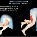
Provocative Examination Maneuvers:
-
Slide Test: Passive hip flexion with knee extension to stretch the musculotendinous complex and reproduce pain at the injury site.
-
Prone Knee Flexion Strength Testing: Tested against resistance at 90*, 45*, and terminal 10* of knee flexion. Pain/weakness at terminal 10* is typically the last deficit to resolve prior to clearance for maximal-velocity sprinting.
-
Reverse Plank Test (Norwig Test): The supine athlete bridges into a reverse plank on the forearms and lifts one leg, transferring eccentric load onto the symptomatic limb. Inability to hold or reproduction of pain indicates incomplete healing and precludes return to play.
-
3. Diagnostic Imaging & Classification
-
Magnetic Resonance Imaging (MRI): Differentiates partial strains, intramuscle belly tearing, and complete bony/tendinous avulsions, while assessing retraction and proximity to the adjacent sciatic nerve.
-
British Athletics Muscle Injury Classification (BAMIC):
-
Grade 0: Normal MRI with clinical focal symptoms (exercise-induced soreness).
-
Grade 1: Small-area myofascial disruption (<10% cross-sectional area [CSA], <5cm length).
-
Grade 2: Moderate strain (10%–50% CSA, 5–10 cm length).
-
Grade 3: Extensive tear (>50% CSA, >75% disruption, or >10cm length).
-
Grade 4: Complete disruption/avulsion of the tendon.
-
Suffix Modifiers: Categorized anatomically as a (myofascial/peripheral), b (musculotendinous junction), or c (intratendinous).
-
4. Proximal Hamstring Tears: Operative vs. Non-Operative Management
-
Complete Proximal Tendon Avulsions:
-
Surgical Indications: Complete 3-tendon avulsions or 2-tendon tears with >2 cm of retraction in active, healthy individuals.
-
Timing: Acute repair within 4 weeks (1 month) of injury yields superior outcomes, functional strength recovery, higher return-to-sport rates (~80%), and fewer sciatic nerve complications compared to delayed repair.
-
Chronic Tears (>4weeks): Complicated by tendon retraction, dense sciatic nerve perineural scarring, and muscle atrophy. While chronic repair outperforms non-operative neglect, it carries higher failure rates, incomplete strength return, and risk of iatrogenic nerve injury.
-
Fixation: Suture anchor fixation to the lateral aspect of the ischial tuberosity (commonly employing 3 or more high-strength suture anchors in a modified Mason-Allen pattern).
-
-
Partial Proximal Tears:
-
Visualized on MRI as the characteristic “sickle sign.”
-
Managed non-operatively initially: activity modification, anti-inflammatory measures, structured rehabilitation, and blood flow restriction (BFR) therapy.
-
Delayed surgical repair of recalcitrant partial tears yields equivalent functional outcomes to early repair, allowing a full trial of conservative therapy without compromising long-term outcomes.
-
-
Role of Biologics (PRP):
-
Leukocyte-poor PRP (LP-PRP) is preferred within muscle bellies to avoid inflammatory lysis; leukocyte-rich PRP (LR-PRP) may be considered for chronic proximal tendinopathies.
-
Ultrasound-guided injections within 24 to 48 hours for Grade 2 strains have demonstrated approximately a 1-game faster return to competition in elite cohorts.
-
5. Distal Hamstring & Musculotendinous Junction Injuries
-
Distal Biceps Femoris Avulsions:
-
Frequently associated with multi-ligament knee injuries and posterolateral corner (PLC) trauma.
-
Complete avulsions warrant anatomical surgical repair back to the fibular head. Care must be taken to dissect and mobilize the common peroneal nerve. Chronic contracted tears may necessitate allograft reconstruction.
-
-
The “T-Zone” Injury:
-
Represents the complex myotendinous junction where the long and short heads of the biceps femoris converge distally (comprising ~35% of distal strains).
-
Exhibits prolonged healing times, high recurrence rates, and delayed return to play (>4–6 weeks). Surgical excision/repair has been documented in recalcitrant, elite athletes to resect the painful fibrous scar nidus.
-
-
Distal Medial Tears (Semitendinosus / Semimembranosus):
-
Isolated complete distal avulsions are uncommon. Most manage conservatively; recalcitrant, painful scarred remnants unresponsive to therapy have occasionally been treated by tenotomy/resection.
-
Master Revision Summary
-
Anatomy: Semimembranosus originates highest and laterally on the ischial tuberosity; semitendinosus and biceps femoris originate as a conjoint tendon more distally and medially.
-
Common Injured Sites: Long head of biceps femoris (take-off phase, eccentric stress) is most frequent; distal biceps “T-zone” involves high recurrence and protracted recovery.
-
Clinical Provocative Tests: Point of maximal tenderness (PMT), slide test, prone knee flexion at terminal $10^\circ$, and the reverse plank (Norwig) test.
-
Proximal Avulsion Rule: Complete tears or 2 retracted tendons (>2cm) warrant acute suture anchor fixation within 4 weeks to avoid sciatic scarring and muscle retraction.
-
Partial Proximal Tears: Trial conservative rehabilitation; delayed repair does not negatively compromise clinical outcome.
-
BAMIC Classification: Stratifies injuries into Grades 0 to 4 with anatomical suffixes: a (myofascial), b (musculotendinous), and c (intratendinous).
-
Return-to-Play Milestones: Absence of pain on the reverse plank test, full strength at terminal 10* of prone knee flexion, and restoration of symmetric high-speed GPS running metrics.
Leave a Reply