
Courtesy: Prof Venu Kavarthapu, King’s College, London, UK
Femoroacetabular Impingement: Diagnosis, Imaging and Hip Arthroscopy
Introduction
Femoroacetabular impingement is an important cause of hip and groin pain in young and physically active adults. It results from abnormal contact between the proximal femur and acetabulum during hip movement.
Repeated mechanical conflict may damage the acetabular labrum and adjacent articular cartilage. In susceptible patients, this can contribute to progressive hip degeneration.
The diagnosis should not be based on radiographic morphology alone. Femoroacetabular impingement syndrome requires appropriate symptoms and clinical findings together with imaging evidence of a relevant morphological abnormality.
Hip Pain in Young Adults
Hip pain in younger patients can arise from:
Extra-articular causes
- Tendinopathy
- Muscle injury
- Greater trochanteric pain syndrome
- Iliopsoas disorders
- Proximal hamstring pathology
- Athletic pubalgia
- Deep gluteal disorders
- Sacroiliac and lumbar pathology
Intra-articular causes
- Femoroacetabular impingement
- Acetabular labral pathology
- Developmental dysplasia of the hip
- Hip instability
- Chondral injury
- Osteonecrosis
- Stress fracture
- Early osteoarthritis
- Synovial and other intra-articular disorders
Femoroacetabular impingement has become an increasingly recognized cause of symptomatic hip pathology in young adults.
What Is Femoroacetabular Impingement?
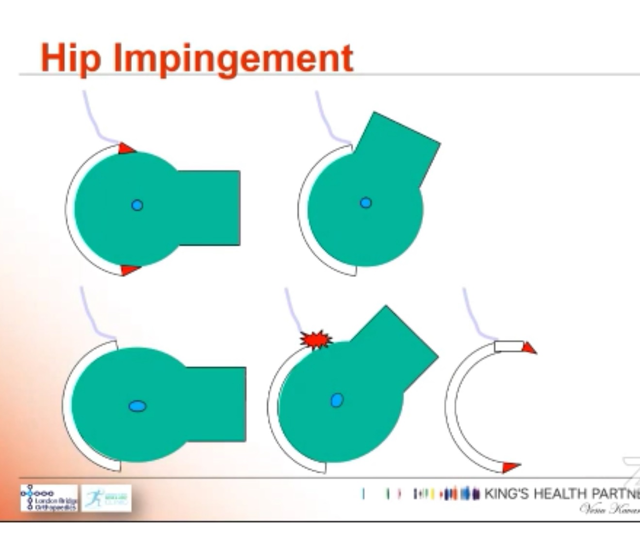
Femoroacetabular impingement is characterized by abnormal contact between the proximal femur and acetabular rim, particularly during flexion and rotation.
The two principal morphological patterns are:
- Cam morphology
- Pincer morphology
Many patients have a combined cam and pincer morphology.
Importantly, cam or pincer morphology may be present in asymptomatic individuals. Therefore:
Morphology alone does not equal femoroacetabular impingement syndrome.
Cam Morphology
Cam morphology represents an abnormal loss of the normal concavity at the femoral head-neck junction.
Instead of a spherical femoral head smoothly transitioning into the femoral neck, there is an aspherical bony prominence.
Mechanism
During hip flexion and rotation:
- The aspherical femoral head-neck junction enters the acetabulum.
- The abnormal prominence produces increased contact with the acetabular rim.
- Repetitive loading may produce:
- Labral injury
- Chondrolabral separation
- Acetabular cartilage damage
- Progressive degenerative changes
Cam morphology is particularly relevant in young athletic individuals and may be associated with activities involving repetitive loading of the hip during adolescence.
Pincer Morphology
Pincer morphology results from excessive acetabular coverage or abnormal acetabular orientation.
The acetabular rim contacts the femoral head-neck region prematurely during hip movement.
Causes include
- Focal anterior acetabular overcoverage
- Excessive global acetabular coverage
- Acetabular retroversion
- Coxa profunda
- Protrusio acetabuli
- Other abnormalities of acetabular orientation
Pincer morphology can also produce:
- Labral compression
- Labral degeneration
- Chondral injury
- Reduced hip range of motion
Combined Cam and Pincer Morphology
Many symptomatic patients have both femoral and acetabular morphological abnormalities.
The clinical problem therefore represents an interaction between:
Femoral morphology + acetabular morphology + hip motion + soft-tissue response + patient activity
rather than a single radiographic abnormality.
Clinical Presentation
The typical patient is a young or middle-aged adult, frequently physically active.
Pain
Pain is commonly:
- Anterior
- Groin
- Deep within the hip
- Occasionally lateral or buttock-related
Patients may describe a deep, poorly localized hip pain rather than pain precisely over the joint.
Activity-related symptoms
Symptoms may initially occur during:
- Running
- Squatting
- Pivoting
- Sports
- Cycling
- Repetitive hip flexion
- High-impact activity
As the condition progresses, pain may occur during ordinary activities such as:
- Getting into or out of a car
- Rising from a low chair
- Sitting for prolonged periods
- Climbing stairs
- Putting on footwear
Advanced disease may produce persistent pain and secondary osteoarthritis.
Clinical Examination
A complete hip examination should be performed rather than relying on a single provocative test.
Examination sequence
Standing
- Gait
- Pelvic alignment
- Limb-length assessment when indicated
- Functional movements
Screening examination
- Lumbar spine
- Sacroiliac joint
- Knee
- Neurological examination when appropriate
Supine examination
Assess:
- Hip flexion
- Extension
- Abduction
- Adduction
- Internal rotation
- External rotation
- Range asymmetry
- Pain reproduction
Hip Impingement Test
The commonly used provocative maneuver is the:
Flexion–Adduction–Internal Rotation Test
The hip is placed into:
Flexion + adduction + internal rotation
This brings the femoral head-neck junction toward the acetabular rim.
A positive test is suggested by:
- Reproduction of the patient’s characteristic pain
- Reduced range of motion
- Particularly restricted internal rotation
However, the test is not specific enough to establish the diagnosis by itself.
Clinical findings must be interpreted alongside imaging and the overall clinical picture.
Differential Diagnosis
Important alternative diagnoses include:
- Developmental dysplasia of the hip
- Hip osteoarthritis
- Acetabular labral tear
- Iliopsoas tendinopathy
- Greater trochanteric pain syndrome
- Proximal hamstring pathology
- Sacroiliac joint disease
- Lumbar radiculopathy
- Stress fracture
- Osteonecrosis
- Deep gluteal syndrome
- Ischiofemoral impingement
- Athletic pubalgia
- Inflammatory arthropathy
- Infection
- Tumour
Imaging
Plain Radiographs
Radiographs remain the initial imaging investigation in most patients.
Useful views include:
- Anteroposterior pelvis
- Dunn view
- Cross-table lateral or other lateral projection
- Additional acetabular orientation views when indicated
Radiographs assess:
- Cam morphology
- Acetabular coverage
- Acetabular orientation
- Joint-space narrowing
- Osteophytes
- Subchondral cysts
- Osteoarthritis
- Other structural abnormalities
Radiographic Assessment of Cam Morphology
Alpha Angle
The alpha angle quantifies femoral head-neck asphericity.
A larger alpha angle indicates greater loss of normal head-neck concavity.
However, there is no single universally accepted alpha-angle cutoff that independently establishes symptomatic femoroacetabular impingement. Thresholds vary according to radiographic projection, measurement technique and clinical context.
An increased alpha angle should therefore be interpreted together with:
- Symptoms
- Physical examination
- Other imaging findings
- Cartilage and labral pathology
Assessment of Pincer Morphology
Acetabular coverage can be assessed using the lateral centre-edge angle.
Broadly:
- Reduced coverage suggests dysplasia.
- Increased coverage may indicate acetabular overcoverage.
Other radiographic signs include:
- Crossover sign
- Posterior wall sign
- Ischial spine sign
- Coxa profunda
- Protrusio acetabuli
- Abnormal acetabular inclination
An increased centre-edge angle does not automatically establish pincer syndrome, because acetabular morphology varies substantially and radiographic measurements can be affected by pelvic positioning.
Computed Tomography
Computed tomography is particularly useful when detailed three-dimensional assessment is required.
It can demonstrate:
- Cam morphology
- Acetabular overcoverage
- Acetabular version
- Femoral version
- Complex femoral morphology
- Extent and location of bony deformity
Three-dimensional reconstructions can be particularly helpful for preoperative planning.
Magnetic Resonance Imaging
Magnetic resonance imaging evaluates both morphology and associated soft-tissue injury.
It can demonstrate:
- Labral tears
- Chondral lesions
- Chondrolabral separation
- Subchondral bone changes
- Cartilage delamination
- Femoral head-neck morphology
- Associated intra-articular pathology
Magnetic resonance arthrography may improve visualization of certain labral lesions, although high-quality modern conventional magnetic resonance imaging can also provide excellent assessment.
Diagnostic Injection
An image-guided intra-articular injection can be valuable when the pain generator is uncertain.
A local anaesthetic injection may help determine whether symptoms originate from the hip joint.
It is particularly useful when:
- Symptoms are poorly localized
- There is concurrent lumbar or sacroiliac pathology
- Clinical examination is equivocal
- Imaging abnormalities may be incidental
- Several potential pain generators are present
A strong temporary response supports an intra-articular pain source, but the test should not be interpreted in isolation.
Management
Management should be individualized.
The major options are:
- Education and activity modification
- Structured rehabilitation
- Analgesic medication when appropriate
- Hip arthroscopy
- Selected open procedures
Non-operative Management
Initial treatment is generally conservative, particularly when symptoms are mild or the diagnosis is uncertain.
Components
- Patient education
- Activity modification
- Avoidance of provocative positions
- Modification of high-impact activities
- Strengthening of hip and trunk musculature
- Movement-control training
- Restoration of functional hip range
- Gradual return to sport
- Appropriate analgesia
Physiotherapy should be structured rather than consisting simply of passive treatment.
Hip Arthroscopy Versus Physiotherapy
The evidence supports both non-operative and operative treatment, with patient selection being important.
The United Kingdom Femoroacetabular Impingement Trial randomized 222 patients with symptomatic femoroacetabular impingement to arthroscopic surgery or physiotherapy and activity modification. At 8 months, arthroscopy produced a greater improvement in the Hip Outcome Score activities-of-daily-living domain.
Therefore:
Hip arthroscopy can provide superior short-term symptom and functional improvement in appropriately selected symptomatic patients, but it is not an indication for every patient with cam or pincer morphology.
Indications for Hip Arthroscopy
Consider surgery when there is:
- Persistent symptomatic femoroacetabular impingement syndrome
- Failure of an appropriate period of conservative treatment
- Clinically relevant cam and/or pincer morphology
- Compatible labral or chondral pathology
- Persistent functional limitation
- Minimal or early osteoarthritis
- Reasonable joint-preservation potential
Patient expectations and activity requirements should also be considered.
Goals of Hip Arthroscopy
The objectives are to:
- Correct pathological femoral morphology
- Correct clinically significant acetabular overcoverage
- Preserve or restore the labrum
- Treat chondral pathology when appropriate
- Restore satisfactory hip mechanics
- Improve pain and function
Arthroscopic Procedures
Depending on pathology, procedures may include:
Cam correction
- Femoral osteoplasty
- Restoration of the femoral head-neck offset
Pincer correction
- Acetabular rim trimming when appropriate
- Correction of focal overcoverage
Labral pathology
- Labral repair
- Selective debridement in appropriately selected cases
- Labral reconstruction when repair is not feasible
Chondral pathology
- Chondroplasty
- Treatment of unstable cartilage
- Marrow-stimulation procedures in selected lesions
Other pathology such as loose bodies or ligamentum teres abnormalities may also be addressed when clinically relevant.
Hip Arthroscopy: Basic Principles
Hip arthroscopy can be performed in either:
- Supine position
- Lateral decubitus position
The essential principle is controlled distraction of the hip to safely access the central compartment.
Fluoroscopy is commonly used during portal establishment.
Because the hip is a deep joint, long instruments and arthroscopes are required.
A 70-degree arthroscope is frequently used, with other viewing angles selected according to the surgeon’s requirements.
Central and Peripheral Compartments
Hip arthroscopy can broadly be divided into:
Central compartment
The space within the acetabular labrum.
Access generally requires hip distraction.
Peripheral compartment
The region outside the labrum but within the capsule.
The peripheral compartment can be accessed without maintaining distraction by manipulating the hip into appropriate positions.
Portal Establishment
Several portal configurations are available.
Common portals include:
- Anterolateral portal
- Mid-anterior portal
- Distal anterolateral accessory portal
- Posterolateral and other accessory portals when required
Portal placement must account for:
- Neurovascular structures
- Labrum
- Articular cartilage
- Capsule
- Femoral head
- Acetabular anatomy
Capsulotomy
Capsulotomy can improve visualization and instrument maneuverability.
It permits:
- Better visualization of the peripheral compartment
- Improved access to the femoral head-neck junction
- Instrument triangulation
- Dynamic assessment of impingement
Capsular management is important because excessive capsular disruption can contribute to postoperative instability, particularly in patients with borderline dysplasia or generalized laxity.
Dynamic Assessment of Cam Morphology
Dynamic assessment is an important part of femoral osteoplasty.
The hip is moved through relevant ranges of:
- Flexion
- Internal rotation
- External rotation
- Abduction
- Adduction
The surgeon identifies the area of pathological contact and maps the cam lesion.
Bone is then progressively resected while maintaining awareness of:
- Femoral neck integrity
- Head-neck offset
- Retained structural strength
- Adjacent cartilage
- Labral relationship
Dynamic reassessment is performed after correction to ensure that clinically relevant impingement has been adequately addressed.
Labral Repair
When the labrum is detached but salvageable:
- The acetabular rim is prepared.
- Suture anchors are positioned.
- Sutures are passed through the labrum.
- The labrum is restored to its anatomical position.
- The repair is tensioned appropriately.
Preservation and repair of the native labrum are generally preferred when the tissue is viable and repairable.
Chondral Injury
Femoroacetabular impingement may produce:
- Focal cartilage damage
- Chondrolabral separation
- Delamination
- Full-thickness cartilage defects
Treatment depends on:
- Size
- Depth
- Location
- Stability
- Patient age
- Degree of osteoarthritis
Treatment may include:
- Chondroplasty
- Stabilization of unstable cartilage
- Marrow stimulation in selected focal defects
- Other cartilage restoration procedures in carefully selected patients
Advanced cartilage damage is an important negative prognostic factor.
Open Surgical Options
Hip arthroscopy is now the predominant joint-preserving surgical approach for most suitable cases.
However, open surgery remains relevant for selected patients with:
- Severe or complex deformity
- Extensive cam morphology not amenable to arthroscopic correction
- Complex acetabular abnormalities
- Complex femoral head deformity
- Certain rotational abnormalities
- Conditions requiring surgical hip dislocation
Surgical hip dislocation provides extensive exposure but carries greater surgical morbidity and is therefore not routinely required for uncomplicated femoroacetabular impingement.
Periacetabular Osteotomy and Acetabular Reorientation
Not every acetabular abnormality should be treated by rim trimming.
If the primary problem is acetabular orientation or structural dysplasia, acetabular reorientation procedures may be more appropriate.
A periacetabular osteotomy may be considered in selected patients with symptomatic structural acetabular dysplasia.
In selected cases of acetabular retroversion with otherwise suitable anatomy, reorientation rather than simple rim resection may be considered.
Postoperative Rehabilitation
Rehabilitation depends on:
- Extent of femoral osteoplasty
- Labral repair
- Cartilage procedure
- Capsular repair
- Bone resection
- Patient characteristics
- Surgeon-specific protocol
Early rehabilitation generally emphasizes:
- Controlled range of motion
- Restoration of gait
- Muscle activation
- Progressive strengthening
- Avoidance of excessive loading during tissue healing
Weight-bearing restrictions vary. More restrictive protocols may be used after extensive labral repair or cartilage procedures.
Return to running and high-level sporting activity is gradual and should be based on:
- Pain
- Range of motion
- Strength
- Neuromuscular control
- Functional testing
- Sport-specific requirements
Complications of Hip Arthroscopy
Potential complications include:
Traction-related
- Pudendal nerve neuropraxia
- Perineal soft-tissue injury
- Sciatic or other nerve symptoms
- Temporary sensory disturbance
Risk is influenced by traction force and duration.
Surgical complications
- Iatrogenic cartilage injury
- Labral injury
- Inadequate correction
- Over-resection
- Femoral neck fracture, particularly after excessive osteoplasty
- Infection
- Heterotopic ossification
- Persistent pain
- Capsular instability
- Recurrent or residual impingement
- Need for revision surgery
- Progression of osteoarthritis
- Conversion to total hip arthroplasty
Residual or Recurrent Impingement
Persistent symptoms after surgery may result from:
- Incomplete cam correction
- Incomplete pincer correction
- Incorrect diagnosis
- Labral pathology
- Cartilage damage
- Hip instability
- Extra-articular pathology
True regrowth of a correctly resected cam lesion is not the usual explanation for postoperative recurrence. Residual morphology or inadequate initial correction is often more relevant.
Prognostic Factors
Factors associated with poorer outcomes include:
- Established osteoarthritis
- Advanced cartilage damage
- Older age
- Reduced joint space
- Significant preoperative degenerative changes
- Severe structural abnormalities
- Persistent symptoms from alternative pain generators
Systematic reviews have consistently identified increasing age and greater joint degeneration as important predictors of less favorable outcomes after hip arthroscopy.
Does Hip Arthroscopy Prevent Osteoarthritis?
This remains an important and evolving question.
It is biologically plausible that correcting pathological mechanics could reduce subsequent cartilage degeneration. However, it should not be presented to patients as a proven method of preventing osteoarthritis.
Recent evidence is encouraging but heterogeneous. A 2025 systematic review found evidence suggesting less radiographic osteoarthritis progression after arthroscopy in some comparative studies, but no statistically significant reduction in conversion to total hip arthroplasty and substantial limitations in the available evidence.
Therefore:
The established indication for hip arthroscopy is treatment of symptomatic femoroacetabular impingement syndrome, not proven prevention of future osteoarthritis.
Long-Term Outcomes
Hip arthroscopy generally produces meaningful improvement in pain and function in appropriately selected patients.
However, outcomes are strongly influenced by:
- Patient selection
- Age
- Cartilage status
- Pre-existing osteoarthritis
- Labral status
- Severity of deformity
Long-term studies demonstrate that some patients eventually require revision surgery or total hip arthroplasty, particularly when degenerative changes are already present.
Leave a Reply