Courtesy: Kevin R Wembridge, Consultant Orthopaedic Surgeon, UK
Sequence of Examination
-
Examination of footwear
-
Gait
-
Inspection
-
Palpation
-
Movements
-
Measurements
-
Neurovascular examination
-
Special tests
1. Examination of Footwear
-
Normal wear
-
Outer side of heel
-
Centre of the sole
-
-
Cavus foot
-
Excess wear on the outer side of heel and lateral border of sole
-
-
Flat foot
-
Excess wear on inner side of heel and medial border of sole
-
-
Bulging of medial footwear wall
-
Suggests everted or flat foot
-
-
Bulging of toe box
-
Suggests claw toes or hammer toes
-
2. Gait Examination
-
Normal gait:
-
Plantigrade feet
-
Equal weight-bearing on heel and forefoot
-
-
Abnormal gait patterns:
-
Antalgic gait
-
High-stepping gait (foot drop)
-
Walking on lateral border of foot (congenital talipes equinovarus)
-
Heel walking: calcaneus deformity
-
Forefoot walking: equinus deformity
-
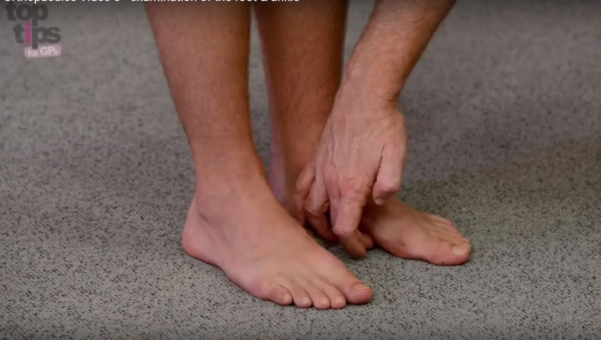
3. Inspection
Inspection should be performed from anterior, lateral, posterior, medial, and plantar aspects.
Anterior Aspect
-
Alignment:
-
Great toe: hallux valgus or hallux varus
-
Lesser toes: claw toes, hammer toes, mallet toes
-
Relationship between forefoot, midfoot, and hindfoot
-
-
Skin condition:
-
Discoloration
-
Ulcers
-
Dilated veins
-
Edema
-
-
Tendons:
-
Extensor hallucis longus
-
Extensor digitorum longus
-
-
Malleolar relationship:
-
Normally, the lateral malleolus lies posterior and inferior to the medial malleolus
-
Lateral Aspect
-
Lateral malleolus
-
Base of the 5th metatarsal
-
Tendo-Achilles
-
Peroneus brevis tendon
-
Lateral border of the foot
Posterior Aspect
-
Hindfoot alignment:
-
Varus or valgus
-
-
Heel:
-
Size and symmetry
-
Position and pattern
-
-
Toe-raise test:
-
Ask patient to stand on tiptoes
-
Normal response shows inversion of hindfoot and increased medial longitudinal arch height (windlass effect)
-
-
Plantar fat pad
-
Calcaneal tuberosity:
-
Prominence of posterosuperior aspect suggests Haglund deformity (pump bump)
-
-
Retrocalcaneal region:
-
Swelling suggests retrocalcaneal bursitis
-
-
Achilles tendon:
-
Tendinitis
-
Rupture (typically 2 to 6 centimeters proximal to insertion)
-
Localized swelling at malleolar level suggests tendinitis
-
Diffuse swelling along tendon suggests rupture
-
-
Calf muscle:
-
Atrophy compared to opposite side
-
Seen in residual deformity of congenital talipes equinovarus, tibialis anterior rupture, or prolonged immobilization
-
Medial Aspect
-
Medial longitudinal arch:
-
Cavus
-
Planus
-
Rocker-bottom deformity (seen in diabetic foot or improperly treated congenital talipes equinovarus)
-
-
Bony prominences:
-
Medial malleolus
-
Head of first metatarsal
-
Navicular tuberosity (prominent in accessory navicular)
-
Calcaneal tuberosity
-
Plantar Aspect
-
Callosities:
-
Indicate abnormal weight-bearing areas
-
Normally present over metatarsal heads and lateral border
-
-
Painful calluses:
-
Common in claw toes and hammer toes due to metatarsophalangeal joint hyperextension
-
-
Corns
-
Ulcerations
-
Warts or fungal infections
4. Palpation
Anterior Palpation
-
Anterior tibial crest: stress fractures
-
Talar dome: osteochondral defects
-
Navicular: Köhler disease
-
First metatarsophalangeal joint: bunions, gout
-
Second metatarsophalangeal joint: Freiberg infarction
-
Interdigital spaces: Morton neuroma
-
Tendons:
-
Tibialis anterior
-
Extensor hallucis longus
-
Extensor digitorum longus
-
Peroneus tertius
-
Lateral Palpation
-
Lateral malleolus
-
Anterior talofibular ligament
-
Calcaneofibular ligament
-
Peroneal tendons
-
Calcaneus and tuberosity: Sever disease
-
Calcaneocuboid joint
-
Sinus tarsi: subtalar arthritis
-
Fibular shaft: stress fractures
Posterior Palpation
-
Gastrocnemius–soleus complex
-
Achilles tendon:
-
Tenderness, swelling, or palpable gap 2 to 6 centimeters above insertion
-
-
Posterior calcaneal tuberosity:
-
Tender swelling in retrocalcaneal bursitis
-
Medial Palpation
-
Medial malleolus
-
Subcutaneous border of tibia
-
Head of talus (palpable on eversion)
-
Navicular tuberosity:
-
Tender swelling in accessory navicular
-
-
Tendons:
-
Flexor hallucis longus
-
Flexor digitorum longus
-
Tibialis posterior
-
5. Movements
-
Dorsiflexion: 20°
-
Plantar flexion: 50°
-
Eversion: 20°
-
Inversion: 40°
-
Abduction: 5°
-
Adduction: 5°
6. Measurements
Longitudinal Measurements
-
True and apparent limb length
-
Heel length:
-
From tip of medial malleolus vertically to heel
-
-
Foot length:
-
Medial: heel to tip of great toe
-
Lateral: heel to tip of 5th toe
-
Circumferential Measurements
-
Thigh
-
Calf
-
Foot at level of medial longitudinal arch
-
Broadening of ankle measured with calipers suggests inferior tibiofibular diastasis
7. Neurovascular Examination
-
Sensory examination
-
Motor examination
-
Peripheral pulses
-
Capillary refill
-
Signs of neuropathy, especially in diabetic patients
8. Special Tests
-
Silfverskiöld test
-
Thompson test
-
Anterior drawer test of ankle
-
Coleman block test
-
Peroneal tendon instability test
-
Tinel test
-
Talar tilt test
-
Morton test
-
Homan sign
Summary
-
Foot and ankle examination must be systematic and comparative.
-
Footwear and gait provide early diagnostic clues.
-
Inspection and palpation help identify deformities and localized pathology.
-
Special tests assist in diagnosing ligamentous, tendon, and nerve conditions.
-
A complete neurovascular examination is essential, especially in high-risk patients.
Leave a Reply