
Courtesy Dr Randy Bindra, Dr Ashok Shyam, Ortho TV
Essex-Lopresti Injuries: Biomechanics, Diagnosis, and Management
Anatomical & Biomechanical Foundation
-
Forearm rotation is an evolutionary specialization unique to humans.
-
Patients tolerate wrist fusion well. They do not tolerate loss of pronation and supination.
-
Load distribution at the wrist is 80% through the radius and 20% through the ulna.
-
Load shifts across the forearm. It reaches 40% radius and 60% ulna at the elbow level.
-
The central band of the interosseous membrane (IOM) transfers this axial load.
-
Central band fibers originate at the pronator teres insertion.
-
Fibers run distally toward the ulna at an angle of 21 degrees.
-
Simple radial head excision allows roughly 7 mm of proximal radial migration if the IOM is intact.
-
Combined rupture of the IOM and Triangular Fibrocartilage Complex (TFCC) causes 15 mm of proximal migration.
-
Complete migration leads to radiocapitellar impingement, ulnocarpal abutment, and severe wrist pain.
Historical Context & True Pathology
-
Brockman (1931): First described distal radioulnar joint (DRUJ) subluxation after radial head excision.
-
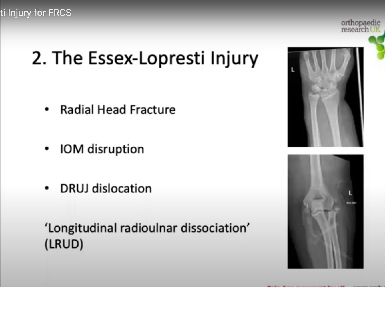
Peter Essex-Lopresti (1951): Reported two post-mortem cases of combined radial head fracture and DRUJ dislocation.
-
Pathoanatomy involves a three-part longitudinal injury:
-
Radial head fracture (often comminuted).
-
Complete rupture of the forearm interosseous membrane (central band).
-
Disruption of the DRUJ with tearing of the TFCC.
-
Diagnostic Approach & Clinical Signs
-
Mechanism: High-energy fall on an outstretched hand (FOOSH).
-
Clinical Red Flag: Proximal elbow pain combined with forearm tenderness and wrist/DRUJ pain.
-
Wrist pain may be absent initially. It manifests prominently if the radial head is mistakenly excised.
-
Radiography: Always obtain wrist X-rays for any comminuted radial head fracture.
-
Frequency: Michael Hausman (New York) noted that 60% of radial head fractures show IOM tears on MRI.
-
Complete Essex-Lopresti dissociations occur in approximately 10% of cases.
Intraoperative Radial Pull Test
-
Perform under fluoroscopy after preparing or removing the broken radial head.
-
Apply longitudinal traction along the radius using a bone reduction clamp.
-
Measure proximal translation relative to the distal ulna on wrist views.
| Proximal Migration Distance | Pathologic Implication | Surgical Action |
| Less than 3 mm | Intact / partially strained IOM | Safe for standard radial head repair |
| 3 mm to 5 mm | TFCC torn; partial IOM compromise | Evaluate DRUJ stability; protect forearm |
| Greater than 6 mm | Complete IOM rupture + TFCC disruption | Definitive radial head replacement mandatory |
Acute Management Algorithm
-
Never excise the radial head: Complete excision leads to chronic proximal migration and joint collapse.
-
Mason Type II: Stable internal screw fixation (tripod screw technique or low-profile plate).
-
Comminuted (Mason Type III/IV): Replace with a metallic radial head arthroplasty as a rigid spacer.
-
Fixation choices:
-
Smooth, loose-fitting stems (Graham King concept) function as rotating spacers.
-
Press-fit, porous-coated stems (Shawn O’Driscoll concept) achieve bony ingrowth.
-
Both approaches report successful outcomes; sizing head diameter and neck height correctly is critical.
-
-
Stabilizing the DRUJ:
-
Transfix the radius and ulna using temporary K-wires in supination.
-
Perform an open or arthroscopic TFCC repair using suture anchors.
-
-
Acute outcomes: Approximately 83% good-to-excellent results with early radial head replacement and DRUJ stabilization.
Chronic Essex-Lopresti Reconstruction
-
Missed diagnosis occurs in roughly 50% of all cases.
-
The Darrach Pitfall: Do not perform a simple Darrach distal ulna excision. The radius will continue migrating proximally, worsening forearm shortening.
-
Failed Radial Head Excision: Insert a metallic radial head implant to restore length, combined with an open TFCC repair.
-
Secondary Capitellar Arthrosis: If cartilage wear prevents placing a metallic head, excise the painful implant and perform formal IOM reconstruction.
IOM Reconstruction: Pronator Teres Transfer Technique
-
Tendon Harvest: Elevate a longitudinal strip of the pronator teres tendon.
-
Leave the distal tendon footprint attached to the radius.
-
Orientation: Pass the tendon strip deep to the extensor tendons toward the ulna at a 21-degree angle.
-
Measure the 21-degree angle intraoperatively using a sterile goniometer.
-
Fixation: Pass the tendon through an ulnar drill hole and secure it under tension.
-
Salvage Option: If multiple reconstructions fail, perform a one-bone forearm fusion in functional semi-pronation.
| Clinical Stage | Radial Component | Forearm & DRUJ Component |
| Acute Presentation | Radial head ORIF or Metallic Arthroplasty | Trans-ulnar K-wire pinning or open TFCC repair |
| Chronic / Missed | Metallic radial head revision | Pronator teres IOM reconstruction (21°) + DRUJ repair |
| End-Stage Salvage | Remove failed hardware | Radioulnar fusion (one-bone forearm) in semi-pronation |

Leave a Reply