
Courtesy: Prof Nabil Ebraheim, University of Toledo, Ohio, USA
Distal Radius Fractures: Types, Biomechanics, and Management
Colles Fracture
-
Definition: An extra-articular distal radius fracture with dorsal displacement of the distal fragment.
-
Mechanism: Low-energy fall onto an outstretched hand (FOOSH).
-
Demographics: Most common in patients older than 50 years.
-
Deformity: Classic dinner fork deformity caused by dorsal tilt and displacement.
-
TFCC Injury: Associated Triangular Fibrocartilage Complex (TFCC) tears occur in roughly 50% of extra-articular fractures.
-
Cast Failure Predictors:
-
Dorsal comminution exceeding 33% to 50% of the dorsal cortex precludes stable cast treatment.
-
More dorsal comminution correlates directly with secondary displacement in a cast.
-
Extension of the fracture line into the distal radioulnar joint (DRUJ) carries the worst prognosis.
-
Smith Fracture (Reverse Colles)
-
Definition: A predominantly extra-articular distal radius fracture with volar (palmar) displacement.
-
Mechanism: Fall onto a flexed wrist.
-
Deformity: Classic garden spade deformity.
-
Treatment: Open reduction and internal fixation (ORIF) via a volar approach using a buttress or locking plate.
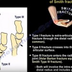
Thomas Classification of Smith Fractures
| Type | Fracture Pattern | Key Anatomical Features |
| Type 1 | Extra-articular transverse | Most common subtype; classic reverse Colles |
| Type 2 | Intra-articular oblique | Fracture line enters the dorsal articular surface |
| Type 3 | Intra-articular oblique | Crosses into radiocarpal joint; identical to a Volar Barton fracture |
Die-Punch Fracture
-
Definition: A depressed, intra-articular fracture of the lunate fossa of the distal radius.
-
Mechanism: Pure axial load transmitted directly through the lunate bone.
-
Clinical Red Flag: High incidence of occult carpal bone dissociation.
-
Assessment: Requires careful evaluation of articular step-off and depression.
Barton Fracture (Fracture-Dislocation)
-
Definition: An intra-articular fracture-subluxation or fracture-dislocation of the radiocarpal joint.
-
Mechanism: Fall onto an extended, pronated wrist with significant shear force.
-
Pathoanatomy: Strong radiocarpal ligaments remain intact. They avulse the articular rim, causing the carpus to dislocate along with the bone fragment.
Volar Barton vs. Dorsal Barton
| Parameter | Volar Barton (Most Common) | Dorsal Barton |
| Displacement | Volar rim avulsion with palmar carpal dislocation | Dorsal rim avulsion with dorsal carpal dislocation |
| Classification Overlap | Equivalent to Smith Type 3 | Often grouped with intra-articular dorsal shear injuries |
| Fragment Size | Variable, typically substantial | Often smaller than volar fragments |
| Surgical Approach | Volar (Henry) approach with volar buttress plate | Dorsal approach with dorsal buttress plate |
Chauffeur Fracture (Hutchinson Fracture)
-
Definition: An isolated intra-articular fracture of the radial styloid process.
-
Mechanism: Direct compression or shear of the scaphoid bone against the radial styloid.
-
Associated Pathology: High risk of concomitant scapholunate dissociation.
-
Imaging Requirement: Always obtain a supinated wrist view to assess the scapholunate interval.
-
Scapholunate Instability & DISI Deformity:
-
Normal scapholunate angle: 47 degrees (normal range: 30 to 60 degrees).
-
Angle greater than 60 degrees indicates abnormal dorsal intercalated segment instability (DISI).
-
The scaphoid flexes volarly while the lunate extends dorsally.
-
AP radiograph shows widening of the scapholunate space (Terry Thomas sign).
-
-
Treatment: Anatomic reduction with compression lag screws across the radial styloid, followed by scapholunate ligament assessment and stabilization if unstable.
Diagnostic Checklist for Distal Radius Fractures
-
Check for articular surface involvement (dorsal vs. volar rim).
-
Assess the lunate fossa for depressed die-punch components.
-
Evaluate the distal radioulnar joint (DRUJ) for articular step-off or widening.
-
Determine carpal translation and the direction of subluxation or dislocation.
-
Inspect carpal intervals on AP and lateral views to rule out scapholunate or lunotriquetral dissociation.

Leave a Reply