• Skip to main content
  • Skip to secondary menu
  • Skip to primary sidebar
OrthopaedicPrinciples.com

OrthopaedicPrinciples.com

Integrating Principles and Evidence

Integrating Principles and Evidence

  • Home
  • Editorial Board
  • Our Books
    • Evidence Based Orthopaedic Principles
  • Courses
  • Exams
  • Reviews
  • Live Program
  • Contact

Distal Femur Physeal Fractures

Courtesy: Prof Nabil Ebraheim, University of Toledo, Ohio, USA


Distal Femoral Physeal Fractures (Pediatric)


Overview

  • Injury to the distal femoral physis in children
  • One of the most important pediatric knee injuries

Clinical Importance

  • High risk of growth arrest (~50–60%)
  • Risk increases with:
    • Displacement
    • High-energy trauma

Key Clinical Point


  • In children presenting with:
    • Suspected MCL/LCL injury

Rule Out

  • Distal femoral physeal fracture

Anatomy and Growth Facts


Growth Contribution

  • ~70% of femoral growth
  • ~37% of total lower limb growth

Growth Rate

  • ~9 mm/year

Ossification

  • First epiphysis to ossify
  • Last to fuse

Fusion Age

  • Girls: ~14 years
  • Boys: ~16 years

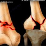
Salter–Harris Classification


Most Common Type

  • Salter-Harris Type II

Key Feature

Thurston–Holland Fragment

  • Triangular metaphyseal fragment
  • Helps identify SH Type II fracture

Pathophysiology


  • Weakest area of physis:
    • Zone of hypertrophy

Special Feature (Distal Femur)

  • Fracture often crosses:
    • Multiple physeal zones

Growth Occurs At

  • Metaphyseal side of physis

Management Principles


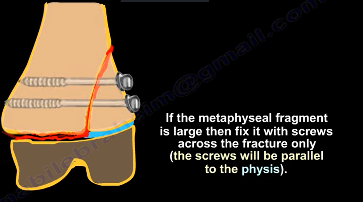
1. Large Metaphyseal Fragment (SH II)


  • Fixation with:
    • Screws

Technical Points

  • Screws:
    • Parallel to physis
    • Avoid physeal damage

2. Small Fragment / Salter I


  • Closed reduction
  • Percutaneous fixation

Implant

  • Smooth K-wires

3. Occult Injury


Scenario

  • Normal X-ray but high suspicion

Next Step

  • MRI or CT scan

When to Suspect

  • Painful knee
  • Instability
  • Pediatric patient

Growth Arrest Patterns


1. Central Arrest


  • Leads to:
    • Limb length discrepancy (LLD)

2. Peripheral Arrest


  • Leads to:
    • Angular deformity

Examples

  • Medial arrest – Genu valgum
  • Lateral arrest – Genu varum

Management of Growth Arrest


< 50% Physis Involved

  • Physeal bar resection
  • Fat interposition

> 50% Physis Involved

  • Epiphysiodesis
  • Growth modulation

Clinical Prediction Examples


  • 12-year-old:
    • ~2 cm shortening expected
  • 13-year-old:
    • ~3 cm predicted – consider intervention

Special Points


  • Minimal shortening if injury occurs:
    • Within 2 years of skeletal maturity

Risk

  • Younger children:
    • Higher likelihood of repeat surgery (8×)

Deformity Prediction Trick


Based on Thurston–Holland Fragment

  • Spike lateral – medial physis injured – Varus deformity
  • Spike medial – lateral physis injured – Valgus deformity

Key Exam Pearls


  • Most common type:
    • Salter-Harris Type II

  • Most serious complication:
    • Growth arrest

  • Best investigation (occult injury):
    • MRI

Treatment Summary

  • Large fragment – Screws
  • Small fragment – K-wires

Critical Clinical Rule

  • Always suspect physeal injury in:
    • “Ligament injuries” in children

Post Views: 1,960

Related Posts

  • Lower trapezius for posterosuperior tears

    Courtesy: Emmanouil Brilakis, Bassam ELhassan, Athens Shoulder Course

  • Fractures of the Distal Femur

    Courtesy: Prof Nabil Ebraheim, University of Toledo, Ohio, USA

  • Distal femur fractures and Nonunion

    ? Courtesy: Prof Nabil Ebraheim, University of Toledo, Ohio, USA DELAYED UNIONS OR NON UNIONS…

Reader Interactions

Leave a Reply

Your email address will not be published. Required fields are marked *

Primary Sidebar

Follow Us

instagram slideshare

Categories

  • -Applied Anatomy
  • -Approaches
  • -Basic Sciences
  • -Cartilage & Meniscus
  • -Classifications
  • -Examination
  • -Foot and Ankle
  • -Foot and Ankle Trauma
  • -FRCS(Tr and Orth) tutorials
  • -Gait
  • -Hand and Wrist
  • -Hand and Wrist Trauma
  • -Hand Infections
  • -Hip and Knee
  • -Hip Preservation
  • -Infections
  • -Joint Reconstruction
  • -Knee Arthroplasty
  • -Knee Preservation
  • -Metabolic Disorders
  • -Oncology
  • -OrthoBiologics
  • -OrthoPlastic
  • -Paediatric Orthopaedics
  • -Paediatric Trauma
  • -Patellofemoral Joint
  • -Pelvis
  • -Peripheral Nerves
  • -Principles
  • -Principles of Surgery
  • -Radiology
  • -Rheumatology
  • -Shoulder and Elbow
  • -Shoulder and Elbow Arthroplasty
  • -Spine Deformity
  • -Spine Oncology
  • -Spine Trauma
  • -Spine, Pelvis & Neurology
  • -Sports Ankle and Foot
  • -Sports Elbow
  • -Sports Knee
  • -Sports Medicine
  • -Sports Medicine Hip
  • -Sports Shoulder
  • -Sports Wrist
  • -Statistics
  • -Technical Tip
  • -Technology in Orth
  • -Trauma
  • -Trauma (Upper Limb)
  • -Trauma Life Support
  • -Trauma Reconstruction
  • Book Shelf
  • Book Shelf Medical
  • Careers
  • Case Studies and Free Papers
  • DNB Ortho
  • Evidence Based Orthopaedic Principles
  • Evidence Based Orthopaedics
  • Exam Corner
  • Fellowships
  • Guest Editor
  • Guest Reviews
  • Image Quiz
  • Instructional Course Lectures
  • Journal Club
  • MCQs
  • Meetings and Courses
  • Multimedia
  • News and Blog
  • Plaster Techniques
  • Podcasts
  • Public Health
  • Rehabilitation
  • Research
  • Shorts and Reels
Copyright@orthopaedicprinciples.com. All right rerserved.