
Courtesy: Prof Nabil Ebraheim, University of Toledo, Ohio, USA
Topic / Overview
Clinical assessment of persistent low back pain in an outpatient setting. Rather than acute post-traumatic pain, this framework focuses on office-based evaluation to stratify simple low back conditions from complicated, sight- or function-threatening spinal disorders.
Background / Problems
-
Clinical Course: Over 90% of patients presenting with acute low back pain improve within 6 to 8 weeks.
-
Target Patient Profile: Patients presenting to an office setting with persistent low back pain that is not resolving, distinct from acute back pain following acute trauma (such as lifting a heavy object or twisting).
-
Diagnostic Challenge: Low back pain can represent either a simple mechanical entity or a complex, multisite spinal disorder. Initial differentiation relies predominantly on targeted history taking, which subsequently directs the focused physical examination and imaging workup.
The Four Essential History Questions
1. Unsteady Gait
-
Question: “Do you have an unsteady gait?” (Specifically querying staggering, instability, or loss of balance).
-
Underlying Pathomechanics: Denotes an upper motor neuron (UMN) lesion, indicating tandem spinal pathology (a second “assault” on the spine at a distant level).
-
Anatomical Localization:
-
Predominantly cervical spine compression (cervical spondylotic myelopathy).
-
Rarely thoracic spinal cord pathology.
-
-
Clinical Implication: Lumbar MRI may account for axial low back pain (e.g., lumbar stenosis), but gait instability mandates an MRI of the cervical spine (or thoracic spine) to rule out myelopathy.
2. Bladder and Bowel Symptoms
-
Question: “Do you have any bladder or bowel symptoms?”
-
Diagnostic Entity: Cauda Equina Syndrome (CES).
-
Symptom Triad: Low back pain, urinary retention, and saddle anesthesia.
-
Pathomechanics: Large central disc herniation compressing the majority of nerve roots within the thecal sac simultaneously, rather than isolated single-root compression.
-
Prognosis & Thresholds:
-
Requires prompt identification; untreated compression leads to permanent sphincter dysfunction.
-
Neurologic recovery of bladder function may remain incomplete despite surgical decompression.
-
Surgical Timing (Speaker-noted controversy): Universal agreement exists on early diagnosis; however, urgency versus emergency timing is debated. A notable clinical threshold is 48 hours—decompression within this window is linked to reasonable outcomes, whereas intervention beyond 48 hours is associated with poorer bladder recovery.
-
3. Relationship of Pain to Activity and Posture
-
Question: “Is the pain related to activity, and is it relieved by rest?”
-
Mechanical / Positional (Spinal Movement):
-
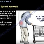
Lumbar Spinal Stenosis (Neurogenic Claudication): Pain is provoked by lumbar extension and alleviated by spinal flexion.
-
Shopping Cart Sign: Relief of symptoms when leaning forward into flexion.
-
-
Metabolic / Muscular (Vascular Claudication):
-
Pain triggered by moving the lower extremity muscles during ambulation and relieved quickly by cessation of walking/standing still.
-
Requires peripheral pulse examination (particularly in elderly cohorts) to rule out vascular disease.
-
Neurogenic and vascular claudication can coexist.
-
-
Non-Mechanical (Constant / Unrelieved by Rest):
-
Pain constant, progressive, and unrelated to activity points toward systemic or non-degenerative etiologies, notably spinal tumor.
-
4. Radiation into the Lower Extremity
-
Question: “Does the pain radiate down into the leg?”
-
Axial vs. Radicular:
-
Axial Pain: Pain localized to the lumbar spine itself; no leg extension.
-
Radicular Pain (Sciatica): Radiation traveling down into the lower extremity.
-
-
Pain Patterns:
-
Radiation Below the Knee: Strongly indicative of lumbar disc herniation.
-
Unilateral Radiation: Suggests focal disc herniation.
-
Bilateral Lower Extremity Radiation: Suggests lumbar spinal stenosis.
-
Indications / Patient Selection for Advanced Imaging
-
Cervical Spine MRI: Indicated when a low back pain patient reports staggering, ataxia, or loss of balance, even if lumbar imaging explains back symptoms.
-
Lumbar Spine MRI:
-
Suspected Cauda Equina Syndrome (emergency/urgent evaluation).
-
Differentiation between lumbar disc herniation and extraspinal sciatic compression (e.g., piriformis syndrome).
-
Persistent, constant pain unrelated to activity to rule out neoplastic lesions.
-
Limitations and Diagnostic Overlaps
-
Normal Physical Examination: A significant proportion of patients with symptomatic lumbar spinal stenosis demonstrate an entirely normal physical examination at rest, making history the primary diagnostic instrument.
-
Spine-Hip Syndrome: Lumbar stenosis and hip osteoarthritis frequently overlap; diagnostic distinction may require a diagnostic hip injection to identify the primary symptom generator.
-
Piriformis Syndrome vs. Lumbar Disc Herniation: Both entities present with identical clinical sciatic distribution; MRI imaging is required to confirm or exclude central/radicular disc pathology.
-
Tandem Stenosis: Lumbar symptoms can mask proximal spinal cord compression in the cervical spine.
Practical Clinical Implications
-
Evaluate peripheral lower-limb pulses routinely in older patients with exertional leg symptoms to differentiate vascular from neurogenic claudication.
-
Any report of balance disturbance in a patient being evaluated for low back pain shifts priority to assessing the cervical spine.
-
Urinary retention in the presence of acute or subacute radicular back pain mandates immediate evaluation for cauda equina compression with an aim for operative intervention within 48 hours.
Key Take-Home Points
-
Prognosis of Back Pain: Over 90% of non-traumatic acute low back pain resolves within 6 to 8 weeks.
-
Four Pillar Queries:
-
Unsteady gait: Signals coexisting cervical/thoracic cord myelopathy.
-
Bowel/bladder dysfunction: Indicates cauda equina syndrome requiring decompression (ideally within 48 hours).
-
Activity relationship: Extension-induced pain indicates stenosis; relief with forward bend is the shopping cart sign; muscle movement vs. rest distinguishes vascular claudication; constant pain warns of tumor.
-
Radiation pattern: Below-the-knee, unilateral pain denotes disc herniation; bilateral involvement indicates spinal canal stenosis.
-


Leave a Reply