Courtesy Dr Kishore Mulpuri , Dr Ashok Shyam, Ortho TV
Developmental Dysplasia of the Hip (DDH)
Introduction
Developmental dysplasia of the hip (DDH) represents a spectrum of abnormalities involving abnormal development of the hip joint.
The spectrum ranges from:
- Mild acetabular dysplasia
- Hip instability
- Subluxation
- Complete dislocation
Early diagnosis and appropriate management are critical to achieving a stable, concentric hip and preventing long-term complications.
Epidemiology and Risk Factors
Important Risk Factors
Major risk factors include:
- Breech presentation
- Positive family history
- Female sex
- First-born child
These patients require careful screening and follow-up.
Pathophysiology
DDH occurs due to abnormal development of:
- The acetabulum
- The femoral head relationship
Persistent instability prevents normal remodeling of the hip joint and may lead to progressive dysplasia and dislocation.
PART 1: Early Diagnosis and Management (Birth–6 Months)
Importance of Early Diagnosis
Early detection allows:
- Non-operative treatment
- Better remodeling potential
- Reduced risk of surgery
- Lower complication rates
Clinical Screening
Barlow Test
The Barlow test evaluates whether the hip can be:
- Dislocated posteriorly
A positive test indicates:
- Hip instability
Ortolani Test
The Ortolani test assesses whether a dislocated hip can be:
- Reduced into the acetabulum
A palpable “clunk” suggests a reducible dislocation.
Imaging
Ultrasound
Ultrasound is the preferred imaging modality in early infancy.
Timing
Best performed at:
- Approximately 6–8 weeks of age
Graf Classification
Ultrasound assessment commonly uses:
- Graf classification
to evaluate acetabular morphology and hip stability.
Plain Radiographs
X-rays become more useful after:
- 4–6 months of age
when femoral head ossification begins to appear.
Management in Early Infancy
Observation
Observation may be appropriate for:
- Mild dysplasia
- Stable hips with improving imaging findings
Pavlik Harness
Indications
Pavlik harness is the standard treatment for:
- Reducible hips
- Early instability
Principle
The harness maintains the hips in:
- Flexion
- Abduction
while allowing motion and promoting concentric reduction.
Important Complication
A known complication is:
- Femoral nerve palsy
which occurs in approximately 5–6% of cases.
Key Clinical Principle
Management decisions should always correlate:
- Clinical examination
- Imaging findings
Neither should be interpreted in isolation.
PART 2: Surgical Management (6 Months–3 Years)
General Principles
The primary goals of treatment are:
- Stable concentric reduction
- Preservation of femoral head vascularity
- Normal hip development
- Prevention of avascular necrosis (AVN)
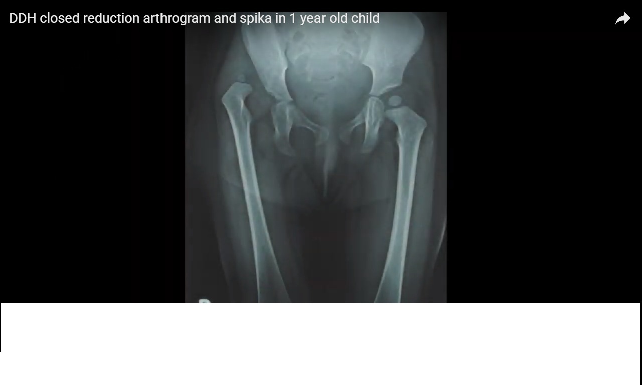
Closed Reduction
Indications
Closed reduction is commonly used in children:
- Approximately 6–15 months old
Arthrogram
An arthrogram is typically performed to assess:
- Concentric reduction
- Obstacles to reduction
Important Principle
The quality of reduction is one of the most important determinants of long-term outcome.
Open Reduction
Indications
Open reduction is indicated for:
- Failed closed reduction
- Irreducible hips
- Severe dislocation
Medial Approach
Advantages
- Minimally invasive
- Direct access to obstructing structures
Limitations
- Limited ability to perform corrective osteotomies
Anterior Approach
Advantages
Provides:
- Wide exposure
- Ability to perform pelvic osteotomy simultaneously
Often preferred in older children.
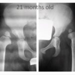
Pelvic Osteotomy
Indications
Pelvic osteotomy is used for:
- Residual acetabular dysplasia
- Inadequate acetabular coverage
It becomes increasingly necessary after:
- Approximately 18 months of age
Femoral Osteotomy
Femoral Shortening
Femoral shortening helps:
- Reduce tension during reduction
- Lower risk of AVN
Derotation Osteotomy
Performed to correct:
- Excessive femoral anteversion
and improve hip stability.
Revision Surgery
Important Considerations
Revision procedures should be avoided whenever possible because they carry:
- Higher complication rates
- Increased stiffness
- Greater AVN risk
Careful primary treatment is essential.
Complications
Potential complications include:
- Avascular necrosis (AVN)
- Residual dysplasia
- Redislocation
- Stiffness
- Limb length discrepancy
- Gait abnormalities
Long-term follow-up is important.
Residual Dysplasia
Residual acetabular dysplasia is one of the most important determinants of:
- Long-term hip function
- Future degenerative arthritis
Continued surveillance during growth is essential.
Key Clinical Pearls
- DDH represents a spectrum from dysplasia to dislocation.
- Early diagnosis significantly improves outcomes.
- Barlow and Ortolani tests are essential neonatal screening tools.
- Ultrasound is the preferred imaging modality in infants.
- Pavlik harness is effective for reducible hips.
- Quality of reduction is more important than the specific surgical technique.
- Forceful reductions increase the risk of AVN.
- Residual dysplasia strongly influences long-term outcome.
Final Take-Home Message
Developmental dysplasia of the hip is a common pediatric hip disorder requiring early recognition and careful management.
Treatment depends on:
- Patient age
- Hip stability
- Severity of dysplasia
The ultimate goal is to achieve and maintain:
- Stable concentric reduction
while minimizing complications such as avascular necrosis and residual dysplasia.
Long-term follow-up is essential to ensure normal hip development and prevent early degenerative arthritis.
Leave a Reply