
Courtesy: Gavin de Kiewiet, Sutherland, UK and Rambam Health Care Campus, Israel
Periacetabular Osteotomy: Principles, Patient Selection and Hip Preservation
Introduction
Successful hip-preservation surgery begins with a detailed understanding of three-dimensional hip anatomy, biomechanics, blood supply and cartilage biology.
Periacetabular osteotomy is primarily a joint-preserving procedure for symptomatic acetabular dysplasia. Rather than creating additional acetabular cartilage, the procedure reorients the existing acetabulum so that the available cartilage is positioned more effectively over the femoral head.
The fundamental objective is to improve:
- Femoral-head coverage
- Joint congruence
- Load distribution
- Hip stability
- Hip biomechanics
- Longevity of the native joint
The operation should therefore be considered as a three-dimensional biomechanical reconstruction, rather than simply an operation to increase radiographic coverage.
Anatomy and Blood Supply: The Foundation of Hip Reconstruction
Knowledge of anatomy is particularly important around the proximal femur and acetabulum because osteotomy changes the relationship between bone, muscles, vessels and joint structures.
Before performing any proximal femoral or pelvic osteotomy, the surgeon should understand:
- Femoral-head blood supply
- Retinacular vessels
- Muscular attachments
- Acetabular anatomy
- Labrum and cartilage
- Neurovascular structures
- Three-dimensional orientation of the pelvis
- Relationship between the femoral head and acetabulum
The precise osteotomy is only one component of reconstruction. Where the cut is made and what structures are preserved are equally important.
Hip Morphology and the Development of Osteoarthritis
Hip morphology varies considerably between individuals.
A patient may have an apparently abnormal radiographic morphology without developing symptoms or osteoarthritis. Conversely, another patient with a similar morphological abnormality may develop significant disease.
Therefore:
Abnormal morphology does not automatically equal symptomatic disease or inevitable osteoarthritis.
Clinical symptoms, physical examination, radiographic morphology, cartilage status and joint congruence must all be interpreted together.
The interaction between morphology and mechanical loading is particularly important. Abnormal coverage or abnormal femoral morphology can concentrate load over a smaller area of articular cartilage, potentially increasing cartilage stress.
Developmental Dysplasia of the Hip
Acetabular dysplasia represents inadequate development or coverage of the femoral head.
Several morphological patterns may be encountered.
Typical features
- Reduced lateral coverage
- Increased acetabular inclination
- Shallow acetabulum
- Increased femoral-head lateralization
- Abnormal acetabular orientation
- Variable anterior deficiency
- Subluxation in more severe cases
Some dysplastic hips demonstrate a relatively broad but insufficiently oriented acetabular roof, whereas others have a steep and short weight-bearing surface.
The combination of poor coverage and subluxation represents a more severe biomechanical problem.
Natural History of Pediatric Dysplasia
The appearance of a pediatric hip on plain radiographs does not always represent the complete functional anatomy of the joint.
In young children, the acetabulum continues to remodel in response to the position and loading of the femoral head.
A concentrically reduced and stable femoral head can stimulate progressive acetabular development.
Conversely:
Persistent instability or eccentric loading
Abnormal acetabular development
Persistent dysplasia and altered load distribution
Therefore, early recognition and appropriate management can take advantage of the remaining growth potential.
Principle of Acetabular Reorientation
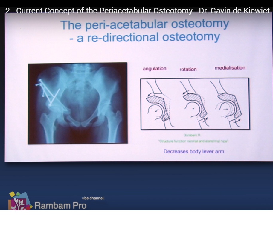
The fundamental concept behind periacetabular osteotomy is redistribution of load across the existing articular cartilage.
The dysplastic acetabulum is not enlarged by the operation.
Instead:
Dysplastic acetabulum
Osteotomy releases acetabular fragment
Three-dimensional reorientation
Improved femoral-head coverage
Larger and better-oriented weight-bearing surface
Reduced focal cartilage loading
Potential preservation of the native hip
This principle is central to modern hip-preservation surgery.
Cartilage Is the Ultimate Target
Improving radiographic measurements is not the final objective.
The true objective is preservation of viable articular cartilage.
If substantial irreversible cartilage damage or advanced osteoarthritis is already present, correcting the acetabular orientation may not restore a durable native hip.
Therefore, cartilage status should influence patient selection.
Assessing Cartilage Before Surgery
Conventional radiographs demonstrate:
- Joint-space preservation
- Osteoarthritis
- Femoral-head shape
- Acetabular morphology
- Subluxation
- Congruence
Advanced imaging can provide additional information about:
- Articular cartilage
- Labrum
- Subchondral bone
- Femoral-head defects
- Femoral morphology
- Acetabular version
Magnetic resonance imaging techniques can be particularly useful when the clinical question concerns cartilage or labral pathology.
However, specialized cartilage imaging should be regarded as an adjunct to clinical assessment, rather than an absolute requirement for every patient.
Age and Cartilage Quality
Young age alone does not guarantee a successful result.
More favorable candidates generally have:
- Symptomatic dysplasia
- Preserved joint space
- Good or acceptable cartilage
- Limited osteoarthritis
- Reasonable joint congruence
- Adequate hip motion
- Correctable acetabular deformity
Advanced osteoarthritis, poor congruence and substantial cartilage damage are associated with inferior outcomes.
Long-term Bernese periacetabular osteotomy studies have consistently identified factors such as older age and pre-existing osteoarthritis as important predictors of failure.
Joint Congruence
Joint congruence is one of the most important determinants of whether reorientation will be beneficial.
A dysplastic hip may appear poorly covered on a standard radiograph but become substantially more congruent when positioned appropriately.
Functional radiographs
Additional views can therefore help determine:
- Whether the femoral head can be concentrically contained
- Whether coverage improves with abduction
- Whether the joint remains congruent after simulated correction
- Whether reorientation is likely to produce an acceptable articulation
This helps distinguish a correctable dysplastic hip from a hip with structural incongruity that may not benefit sufficiently from acetabular reorientation.
The Femur Must Also Be Assessed
Acetabular dysplasia frequently coexists with femoral abnormalities.
These may include:
- Excessive femoral anteversion
- Coxa valga
- Femoral-head deformity
- Femoral-head-neck abnormalities
- Trochanteric abnormalities
- Proximal femoral shortening or deformity
Therefore:
Do not evaluate the acetabulum in isolation.
A technically excellent periacetabular osteotomy can still produce a poor result if a major femoral deformity remains untreated.
Combined acetabular and femoral reconstruction may therefore be necessary in selected patients.
Labrum and the Chondrolabral Junction
The acetabular labrum has important biomechanical functions.
It contributes to:
- Joint sealing
- Fluid pressurization
- Load distribution
- Joint stability
- Protection of the peripheral cartilage
Labral abnormalities are common, including in asymptomatic individuals, particularly with increasing age. Consequently, the mere presence of a labral tear on magnetic resonance imaging does not establish it as the cause of symptoms.
The surgeon must determine whether the labral abnormality is:
- Incidental
- Secondary to dysplasia
- Associated with femoroacetabular impingement
- Associated with cartilage injury
- A major contributor to symptoms
Labral Management During Hip Preservation
The historical approach of simply excising damaged labral tissue has increasingly given way to labral preservation and repair whenever technically appropriate.
Potential options include:
- Labral repair
- Labral reconstruction in selected deficient labra
- Limited debridement in carefully selected cases
- Treatment of the underlying acetabular or femoral abnormality
The key principle is:
Treat the cause of labral injury, not merely the labral tear.
For example, repairing a labral tear without correcting significant dysplasia or femoroacetabular impingement may leave the underlying abnormal mechanics unchanged.
Importantly, evidence does not demonstrate that every labral tear requires simultaneous intervention during periacetabular osteotomy. One clinical study found similar short-term outcomes after periacetabular osteotomy in dysplastic hips with and without labral tears. Treatment should therefore be individualized according to symptoms, morphology and associated pathology.
Cartilage Lesions
Cartilage damage represents a major prognostic concern.
Possible lesions include:
- Focal chondral defects
- Chondral flaps
- Delamination
- Osteochondral defects
- Subchondral cystic change
- Diffuse degenerative cartilage loss
Selected focal defects may be treated with procedures such as:
- Chondroplasty
- Microfracture
- Osteochondral transplantation
- Matrix-based cartilage procedures
- Fixation of suitable cartilage or osteochondral flaps
However, these procedures have variable evidence and should not be interpreted as reliably restoring a biologically normal joint.
The most important intervention remains correction of the abnormal mechanical environment whenever the underlying morphology is correctable.
Femoroacetabular Impingement Must Be Addressed
Dysplastic hips can coexist with femoral or acetabular morphology capable of producing impingement.
Examples include:
- Cam morphology
- Acetabular retroversion
- Focal anterior overcoverage
- Residual deformity following previous surgery
If an impinging deformity is left untreated, postoperative symptoms may persist despite technically successful acetabular reorientation.
Therefore, the final hip should be assessed dynamically for:
- Flexion
- Internal rotation
- Adduction
- Anterior impingement
- Residual femoroacetabular conflict
Selected patients may require concomitant femoral osteochondroplasty or treatment of focal acetabular abnormalities.
Periacetabular Osteotomy
The Bernese periacetabular osteotomy was developed to permit substantial acetabular reorientation while preserving the posterior column.
It has become an established joint-preserving operation for appropriately selected adolescents and adults with symptomatic acetabular dysplasia.
Basic principle
The acetabulum is released through a series of controlled osteotomies.
The mobile fragment is then:
- Rotated
- Tilted
- Translated
until satisfactory three-dimensional coverage and joint congruence are obtained.
Three-Dimensional Reorientation
The acetabulum is oriented obliquely within the pelvis.
Therefore, correction cannot be planned simply by thinking in terms of one radiographic angle.
The surgeon must consider:
- Lateral coverage
- Anterior coverage
- Posterior coverage
- Acetabular inclination
- Acetabular version
- Medialization
- Femoral morphology
- Functional range of motion
Three-dimensional computed tomography can be particularly useful in complex deformity, version abnormalities and combined femoral-acetabular pathology.
Acetabular Retroversion
Periacetabular osteotomy is not restricted to classic lateral acetabular deficiency.
Selected patients may have:
- Global acetabular retroversion
- Focal anterior or superior retroversion
- Abnormal anterior coverage
- Combined dysplasia and version abnormalities
The key question is:
Is the abnormality localized to a small part of the acetabulum, or does the entire acetabular orientation require correction?
A focal rim abnormality may sometimes be managed with limited acetabular correction or arthroscopic treatment.
A global version abnormality may require reorientation of the acetabulum itself.
Importance of Standardized Radiographs
Acetabular measurements are highly dependent on pelvic positioning.
Radiographs should therefore be obtained using a standardized technique.
Variations in:
- Pelvic tilt
- Pelvic rotation
- Beam position
- Patient position
can substantially alter apparent acetabular coverage and version.
Consequently:
An apparently abnormal measurement on a poorly positioned radiograph should not be used in isolation to make a surgical decision.
Radiographic Assessment
Important measurements may include:
- Lateral center-edge angle
- Anterior center-edge angle
- Acetabular index or Tönnis angle
- Femoral-head extrusion
- Acetabular version
- Anterior wall coverage
- Posterior wall relationship
- Shenton line
- Femoral-head sphericity
These measurements should be interpreted collectively.
No single angle completely describes the three-dimensional acetabular morphology.
Imaging the Femoral Head and Neck
Three-dimensional imaging may be particularly useful when planning combined femoral and acetabular correction.
The objective is to identify:
- Cam morphology
- Femoral version
- Femoral-neck orientation
- Femoral-head deformity
- Trochanteric position
- Areas requiring osteochondroplasty
The principle is selective correction:
Remove or correct only the morphology that is demonstrably abnormal and clinically relevant.
Over-resection of the femoral head-neck junction can itself create instability or structural weakness.
Periacetabular Osteotomy for Instability
Some patients develop instability because acetabular deficiency was not recognized or because previous hip-preservation surgery altered the mechanical environment.
Examples include:
- Unrecognized anterior acetabular deficiency
- Iatrogenic instability following hip arthroscopy
- Generalized ligamentous laxity
- Neuromuscular conditions
- Syndromic hip instability
In selected cases, acetabular reorientation can restore coverage and improve stability.
However, the exact cause of instability must be established before performing reconstruction.
Syndromic and Neuromuscular Conditions
Some patients with:
- Down syndrome
- Selected connective-tissue disorders
- Neuromuscular disease
- Other syndromic conditions
may develop progressive hip instability.
The treatment must account for:
- Muscle imbalance
- Ligamentous laxity
- Neurological status
- Bone morphology
- Recurrence risk
Acetabular osteotomy may be appropriate in selected patients, but it should form part of a broader reconstruction strategy rather than being viewed as an isolated solution.
Combined Proximal Femoral Reconstruction
Periacetabular osteotomy may need to be combined with proximal femoral procedures.
Potential indications include:
- Abnormal femoral version
- Coxa valga or vara
- Femoral shortening
- Femoral-head deformity
- Trochanteric overgrowth
- Residual deformity from childhood hip disease
Combined correction should be planned as a single biomechanical reconstruction.
Legg-Calvé-Perthes Disease
Patients with residual deformity following Legg-Calvé-Perthes disease may develop:
- Femoral-head asphericity
- Coxa magna
- Coxa breva
- Trochanteric overgrowth
- Reduced femoral-neck offset
- Acetabular dysplasia
- Femoroacetabular impingement
Treatment may therefore require a combination of:
- Acetabular reorientation
- Femoral osteotomy
- Trochanteric advancement or transfer
- Femoral-head-neck reshaping
- Labral repair
The objective is to restore the best possible relationship between the femoral head and acetabulum.
Cartilage Biology and Joint Preservation
Joint preservation is increasingly viewed as a combination of mechanical and biological management.
Cartilage degeneration involves interactions between:
- Mechanical overload
- Chondrocyte dysfunction
- Inflammatory mediators
- Oxidative stress
- Cellular senescence
- Matrix degradation
These mechanisms are important areas of current research.
However, experimental biological concepts should not be confused with established clinical treatments. At present, correction of pathological joint mechanics remains the central proven principle of hip-preservation surgery.
Patient Selection for Periacetabular Osteotomy
The ideal candidate generally has:
Favorable features
- Symptomatic acetabular dysplasia
- Preserved joint space
- Limited osteoarthritis
- Good or acceptable cartilage
- Reasonable joint congruence
- Correctable deformity
- Adequate hip motion
- Sufficient biological potential for preservation
Less favorable features
- Advanced osteoarthritis
- Severe cartilage loss
- Marked incongruence
- Severe femoral-head deformity
- Very limited range of motion
- Uncorrectable femoral deformity
- Previous extensive surgery
Long-term studies demonstrate that pre-existing osteoarthritis and increasing age are important determinants of poorer survivorship.
Borderline Dysplasia: A Difficult Decision
A patient with mildly reduced acetabular coverage presents a particular challenge.
The decision should not be based on a single measurement.
Consider:
- Symptoms
- Instability
- Lateral coverage
- Anterior coverage
- Acetabular inclination
- Acetabular version
- Femoral version
- Femoral morphology
- Labral pathology
- Cartilage status
- Functional limitations
The critical question is whether the morphology is actually responsible for the patient’s symptoms and whether correction is likely to provide meaningful benefit.
When Should Surgery Be Considered?
Observation may be appropriate for an asymptomatic patient with radiographic dysplasia, depending on age, severity and clinical context.
Surgery becomes more appropriate when there is:
- Persistent symptoms
- Functional limitation
- Mechanical instability
- Correctable dysplasia
- Preserved joint cartilage
- Acceptable joint congruence
- Reasonable expectation of joint preservation
There is no universal requirement to operate on every incidentally discovered dysplastic hip.
Long-Term Outcomes
Periacetabular osteotomy can provide durable pain relief and functional improvement in appropriately selected patients.
However, it is not a guarantee against future osteoarthritis or total hip arthroplasty.
The original Bernese series has demonstrated meaningful long-term native-hip survival, but a substantial proportion of patients eventually require total hip arthroplasty. In the initial 63-patient, 75-hip cohort followed for approximately 30 years, 42 hips had been converted to total hip arthroplasty by the latest follow-up.
This emphasizes an important principle:
Periacetabular osteotomy delays or prevents joint failure in selected patients; it does not make the hip biologically immune to osteoarthritis.
What Happens if the Hip Eventually Fails?
Total hip arthroplasty remains an effective salvage procedure after failed periacetabular osteotomy.
Modern evidence is more reassuring than early reports might suggest.
A 2025 systematic review found that patients undergoing total hip arthroplasty after previous pelvic osteotomy generally had comparable functional outcomes, complication rates and survivorship to patients undergoing primary total hip arthroplasty, although surgery may be technically more demanding, with longer operative time, greater blood loss and more challenging acetabular component positioning.
A separate mid-term study of total hip arthroplasty after periacetabular osteotomy reported substantial functional improvement and approximately 96% survivorship free from revision at 5 and 10 years.
Thus:
Previous periacetabular osteotomy does not preclude successful future total hip arthroplasty.
Important Planning Consideration for Future Arthroplasty
Because periacetabular osteotomy changes acetabular orientation, future arthroplasty requires careful assessment of:
- Acetabular version
- Bone stock
- Hip center
- Component orientation
- Previous screw positions
- Femoral anatomy
- Trochanteric anatomy
- Previous femoral osteotomies
Previous proximal femoral reconstruction can create additional challenges during future arthroplasty.
Contemporary evidence suggests that the principal issue is technical complexity, rather than an inevitable poor arthroplasty outcome.
Causes of Failure After Periacetabular Osteotomy
Failure may result from:
- Poor patient selection
- Advanced osteoarthritis
- Poor cartilage
- Severe incongruence
- Uncorrectable deformity
- Inadequate correction
- Persistent dysplasia
- Persistent instability
- Residual abnormal load distribution
- Excessive correction
- Anterior impingement
- Excessive retroversion
- Reduced range of motion
- Unrecognized femoral deformity
- Excessive anteversion
- Cam morphology
- Coxa valga
- Femoral-head deformity
- Residual intra-articular pathology
- Untreated labral pathology
- Chondral injury
- Femoral-head defects
- Progressive cartilage degeneration
Even an anatomically successful osteotomy cannot reliably reverse established advanced cartilage loss.
Intraoperative Assessment
The operation should not end once the acetabular fragment appears appropriately positioned on fluoroscopy.
The surgeon should assess:
- Hip range of motion
- Anterior impingement
- Flexion
- Internal rotation
- Joint congruence
- Stability
- Femoral-head coverage
- Acetabular version
Dynamic assessment can identify impingement that is not obvious from a single static radiograph.
Practical Principles
Principle 1 — Understand the anatomy
Know the blood supply, muscles, neurovascular structures and three-dimensional pelvic anatomy before making the osteotomy.
Principle 2 — Treat the patient, not the radiograph
Radiographic dysplasia without symptoms does not automatically require surgery.
Principle 3 — Preserve cartilage
The condition of the articular cartilage is one of the most important determinants of long-term success.
Principle 4 — Check congruence
A technically perfect reorientation is of little value if it produces an incongruent articulation.
Principle 5 — Evaluate the femur
Acetabular correction alone may be inadequate when significant femoral deformity is present.
Principle 6 — Preserve the labrum when possible
Treat labral pathology selectively, while correcting the underlying mechanical abnormality.
Principle 7 — Look for impingement after correction
The final hip must have satisfactory motion without creating a new mechanical conflict.
Principle 8 — Think in three dimensions
Coverage, inclination, version, medialization and femoral morphology must be considered together.
Leave a Reply