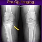
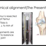
Courtesy: Dr S Macdonald, Ashok Shyam, Ortho TV
Flexion contracture is frequently encountered in advanced osteoarthritis—particularly in populations presenting late. It significantly affects gait mechanics and functional recovery.
Why It Matters
-
Flexion contracture increases quadriceps workload.
-
Bilateral 30° contractures ? ~50% increase in quadriceps effort.
-
Leads to:
-
Energy-inefficient gait
-
Reduced walking endurance
-
Poor postoperative satisfaction if uncorrected
-
Grading of Flexion Contracture
| Grade | Deformity |
|---|---|
| Grade 1 | < 15° |
| Grade 2 | 15°–30° |
| Grade 3 | > 30° |
Grade 2 and 3 deformities are commonly seen in advanced OA.
Pathoanatomy
Structures contributing to flexion deformity:
-
Posterior capsule
-
Posterior cruciate ligament (PCL)
-
Posterior osteophytes (femur and tibia)
-
Hamstrings
-
Gastrocnemius
-
Posterior soft tissue envelope
In inflammatory arthritis:
-
Additional posterior bone loss contributes.
?? Important Principle
Avoid correcting flexion deformity by excessive distal femoral resection alone.
Over-resection leads to:
-
Flexion instability
-
Raised joint line
-
Poor long-term function
Always prioritize soft tissue correction first.
Three-Stage Management Strategy
1?? Preoperative Measures
Limited role, but useful in select cases:
-
Serial manipulation
-
Serial casting
-
Epidural-assisted stretching (especially inflammatory arthritis)
-
Correct hips first if bilateral hip and knee involvement
?? Risk: Supracondylar fracture during aggressive manipulation.
2?? Intraoperative Management (Key Phase)
Step 1: Adequate Exposure
-
Standard approach.
-
Maintain measured bone resection technique.
Step 2: Posterior Osteophyte Removal (Critical Step)
-
Remove femoral and tibial posterior osteophytes completely.
-
Use curved osteotome or rongeur.
-
Work carefully under the MCL.
-
Recreate posterior recess.
This alone often significantly reduces contracture.
Step 3: Posterior Soft Tissue Release
-
Subperiosteal release from posterior femur.
-
Can safely extend deep without vascular compromise if performed correctly.
Step 4: Tibial Slope
-
Minimum ~5° posterior slope recommended.
-
Inadequate slope may perpetuate flexion tightness.
-
Particularly important in CR knees.
Step 5: PCL Management
-
PCL sacrifice often required in moderate-to-severe deformity.
-
CR correction possible in selected mild cases.
-
PS or constrained designs often preferred in severe cases.
Step 6: Posterior Capsulotomy
-
Performed under direct visualization.
-
Can be done in flexion or extension.
-
Use laminar spreaders.
-
Release central posterior capsule carefully.
Almost always required in Grade 2–3 deformities.
Step 7: Gastrocnemius Release (Selective)
-
Consider in severe fixed deformity.
-
Especially when posterior tightness persists.
Step 8: Distal Femoral Recut (Last Resort)
-
If contracture persists:
-
Additional 2 mm distal femoral resection.
-
-
Avoid excessive resection.
-
Preserve joint line integrity.
3?? Postoperative Management
Key Principles
-
No pillow under the knee.
-
Pillow under heel to maintain extension.
-
Night knee immobilizer.
-
Controlled use of CPM.
-
Aggressive extension exercises.
Failure in postoperative extension positioning can undo intraoperative correction.
Correction Expectations
Osteoarthritis
-
Aim for full correction.
-
Up to 10° residual deformity may gradually correct over time.
Rheumatoid Arthritis
-
Full correction not always achievable.
-
Functional improvement more important than absolute zero deformity.
Additional Considerations
Patella Baja
-
Avoid excessive distal femoral resection.
-
Avoid joint line elevation.
-
May consider:
-
Tibial resection adjustments
-
AP femoral sizing modifications
-
Severe Contracture (Extreme Cases)
-
Consider:
-
Medial capsule advancement
-
Quadriceps slide
-
Double-breasting technique
-
Constrained implants if instability present
-
Complications to Watch For
-
Neurovascular injury
-
Wound healing problems
-
Recurrent deformity
-
Ligament instability
-
Posterior subluxation
Careful, stepwise correction minimizes these risks.
Practical Algorithm
-
Remove posterior osteophytes.
-
Posterior capsular release.
-
PCL sacrifice (if needed).
-
Gastrocnemius release (if needed).
-
Reassess extension gap.
-
Additional distal femoral resection only if necessary.
-
Consider constrained implant if instability.
-
Enforce strict postoperative extension protocol.
Final Clinical Pearls
-
Do not rush bone resection.
-
Soft tissue correction is primary.
-
Joint line preservation is critical.
-
Focus is essential in severe deformities.
-
Inadequate attention ? instability disaster.

Leave a Reply