Courtesy: Vinay Kumar Signh, NIMS and EVA Hospital, Jaipur, India
General Examination
-
General build and nutritional status.
-
Presence of pallor, icterus, cyanosis, clubbing, lymphadenopathy, or edema.
-
Vital signs.
-
Height, weight, body mass index, and arm span.
-
Neurocutaneous markers.
-
Secondary sexual characteristics.
-
Evidence of generalized ligamentous laxity.
Local Examination
Local examination of the elbow includes:
-
Inspection
-
Palpation
-
Movements
-
Measurements
-
Special tests
Inspection
-
The elbow should be inspected from the anterior, lateral, medial, and posterior aspects.
-
The patient is asked to place both upper limbs in the anatomical position for comparison.
Attitude of the Limb
-
Observe how the limb is held at rest.
-
Protective flexion posture may suggest pain or injury.
Alignment of the Elbow and Forearm
-
Assess the carrying angle.
Carrying Angle
-
Patient stands with the arm close to the chest and forearm supinated.
-
The angle between the long axis of the arm and the long axis of the forearm represents the carrying angle.
-
Normal values:
-
Males: 5 to 10 degrees
-
Females: 10 to 15 degrees
-
-
Carrying angle cannot be accurately assessed in the presence of a fixed flexion deformity.
Deformities of the Elbow
-
Flexion deformity:
-
Common after elbow trauma or arthritis.
-
-
Cubitus valgus:
-
Seen after lateral condyle fractures and occasionally supracondylar fractures.
-
-
Cubitus varus (gunstock deformity):
-
Classical malunion following supracondylar fracture of the humerus.
-
Anterior Aspect Inspection
-
Inspect the cubital fossae on both sides.
-
Look for:
-
Fullness or swelling, such as myositis ossificans
-
Visible pulsations or prominent veins
-
Sinuses or surgical scars
-
Muscle wasting
-
Lateral Aspect Inspection
-
Observe for swelling and deformity.
-
Assess muscle contour.
-
Identify the mobile wad of three muscles.
-
Inspect biceps and triceps:
-
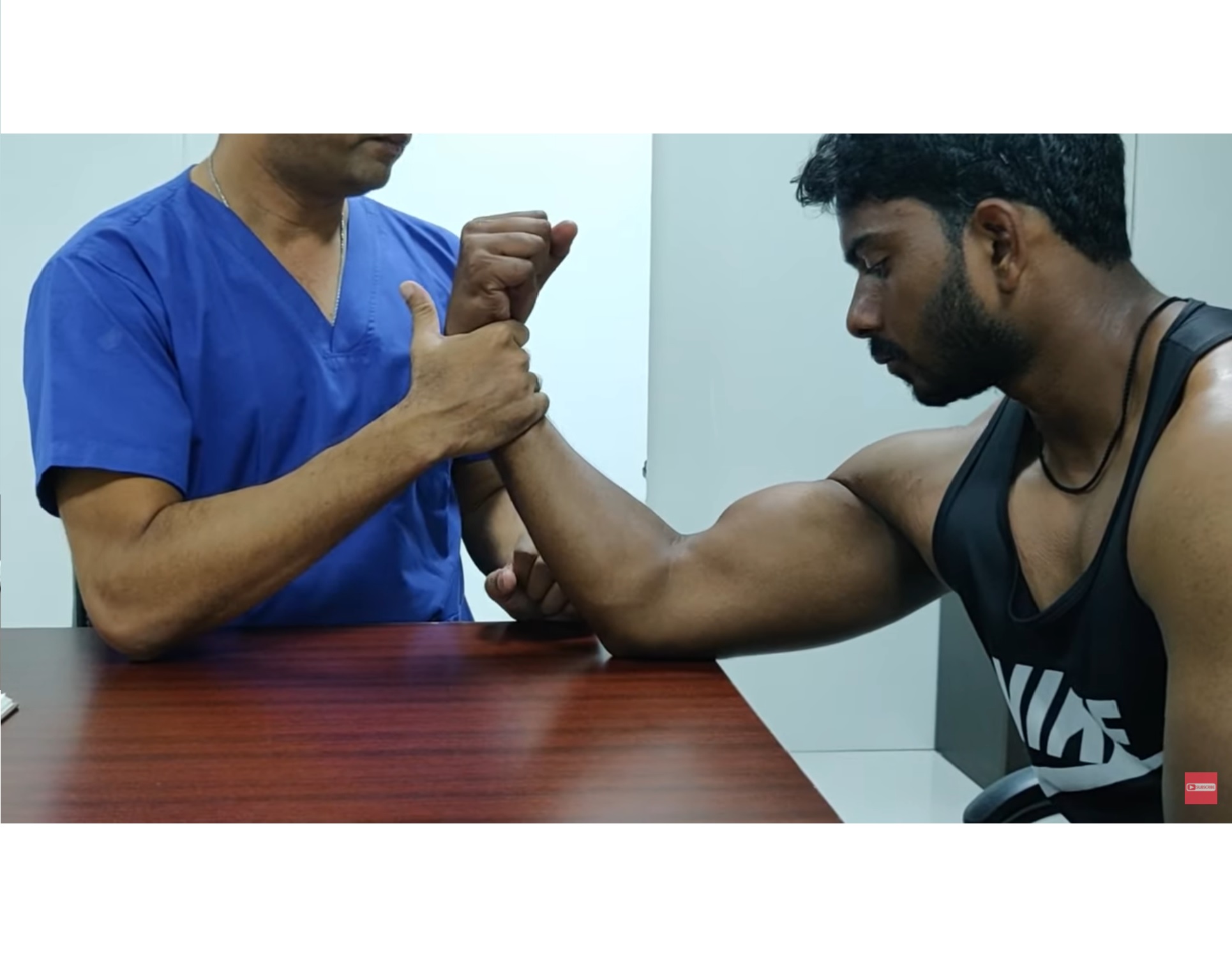
Popeye sign suggests biceps tendon rupture.
-
-
Tenting of triceps:
-
Seen in posterior dislocation of the elbow.
-
-
Look for swelling over the olecranon.
-
Inspect the anconeus triangle for fullness.
Posterior Aspect Inspection
-
Observe:
-
Triceps tendon
-
Olecranon prominence
-
Para-olecranon region
-
Medial and lateral epicondyles
-
-
Look for:
-
Abnormal lumps or swelling, such as in neglected elbow dislocation
-
Muscle wasting
-
Sinuses or scars
-
Prominent veins
-
-
Assess the three-point bony relationship.
Palpation
-
Local rise of temperature.
-
Tenderness.
-
Palpation of bony landmarks:
-
Lateral epicondyle
-
Olecranon
-
Radial head
-
Medial epicondyle
-
Soft Tissue Palpation
-
Anteriorly:
-
Palpate the cubital fossa for swelling or tenderness.
-
Palpate the biceps tendon and lacertus fibrosus during resisted elbow flexion.
-
-
Medially:
-
Palpate the ulnar nerve just posterior to the medial epicondyle.
-
Palpate the epitrochlear lymph node.
-
-
Posteriorly:
-
Palpate for swelling in the para-olecranon region.
-
Palpate the tip of the olecranon.
-
-
Laterally:
-
Palpate the anconeus triangle for effusion or tenderness.
-
Palpate one finger breadth distal to the lateral epicondyle to assess the origin of extensor carpi radialis brevis in lateral epicondylitis.
-
-
Joint lines:
-
Anterior joint line
-
Posterior joint line
-
Radiocapitellar joint
-
Movements of the Elbow
-
Flexion: 0 to 140 degrees
-
Extension: up to 10 degrees of hyperextension
-
Pronation: 0 to 70 degrees
-
Supination: 0 to 85 degrees
Measurements
Upper Limb Length
-
Arm length:
-
Anterolateral border of the acromion to the lateral epicondyle.
-
-
Forearm length:
-
Lateral epicondyle to radial styloid.
-
Hueter Triangle
-
Distance from olecranon to medial epicondyle.
-
Distance from olecranon to lateral epicondyle.
Muscle Wasting
-
Identify the area of maximum wasting.
-
Measure circumferential girth and compare with the opposite side.
Bone Length Measurements
-
Radial length:
-
Lateral epicondyle to radial styloid.
-
-
Ulnar length:
-
Olecranon tip to ulnar styloid.
-
Carrying Angle
-
Measured between the long axis of the arm and the long axis of the forearm.
Special Tests
Elbow Instability
-
Varus stress test
-
Valgus stress test
-
Posterolateral rotatory instability test
-
Chair push-up test
Lateral Epicondylitis (Tennis Elbow)
-
Cozen test
-
Mills test
-
Maudsley test
-
Chair test
Medial Epicondylitis (Golfer’s Elbow)
-
Resisted wrist flexion test
Radial Tunnel Syndrome
-
Long finger extension test
Pronator Syndrome
-
Prolonged resisted pronation test
-
Prolonged resisted elbow flexion test
-
Long finger proximal interphalangeal joint flexion test
Cubital Tunnel Syndrome
-
Tinel sign at the elbow
-
Prolonged elbow flexion test
-
Ulnar nerve compression test
Other Examinations
-
Examination of joints above and below:
-
Shoulder
-
Wrist
-
-
Regional lymph node examination:
-
Cubital fossa
-
Axilla
-
Neurovascular Examination
-
Deep tendon reflexes:
-
Biceps reflex
-
Triceps reflex
-
Supinator reflex
-
-
Motor and sensory examination of:
-
Ulnar nerve
-
Median nerve
-
Radial nerve
-
-
Peripheral pulses:
-
Radial pulse
-
Ulnar pulse
-
Leave a Reply