Courtesy: Prof Nabil Ebraheim, University of Toledo, Ohio, USA
Complications of Talar Neck Fractures
Overview
- Talar neck fractures can result in serious complications affecting:
- Joint function
- Bone viability
Major Complications
- Post-traumatic arthritis
- Avascular necrosis (AVN)
- Malunion
- Nonunion
Post-Traumatic Arthritis
Subtalar Arthritis
- Most common complication
- Incidence:
- ~50–100%
Cause
- Cartilage damage at time of injury
Ankle (Tibiotalar) Arthritis
- Incidence:
- ~30%
Avascular Necrosis (AVN) of Talus
Definition
- Death of bone due to loss of blood supply
Pathophysiology
- Primarily affects:
- Body of talus
- Usually partial involvement, not entire talus
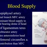
Blood Supply of Talus
Key Vessels
- Artery of tarsal canal (dominant)
- Deltoid branch of posterior tibial artery
- Artery of tarsal sinus
Critical Point
- Deltoid branch may be:
- Only remaining blood supply in displaced fractures
Surgical Importance
- Must be preserved during surgery
Risk Factors for AVN
- Increased fracture displacement
- Severe injury patterns
- Open fractures
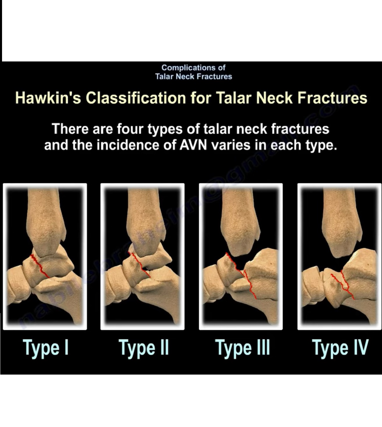
Hawkins Classification & AVN Risk
| Type | Description | AVN Risk |
|---|---|---|
| Type I | Undisplaced | ~10% |
| Type II | Subtalar subluxation/dislocation | ~50% |
| Type III | Subtalar + ankle dislocation | ~90% |
| Type IV | + talonavicular dislocation | ~100% |
Overall AVN Incidence
- ~30%
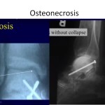
Diagnosis of AVN
Hawkins Sign
Definition
- Subchondral radiolucent line in talar dome
Timing
- Appears at:
- 6–8 weeks
Significance
- Indicates:
- Preserved blood supply
- Good prognosis
Diagnostic Value
- Sensitivity:
- ~100%
- Specificity:
- ~57%
Important Note
- Absence does not confirm AVN
Imaging
MRI
- Detects early AVN
- Shows:
- Reduced T1 signal
Limitation
- May not change treatment
Implant Consideration
- Titanium implants:
- Less MRI artifact than stainless steel
Nonunion
- Incidence:
- ~5%
Definition
- Failure of fracture healing
Malunion
1. Varus Malunion
Incidence
- ~25–30%
Cause
- Medial comminution
Clinical Features
- Hindfoot varus deformity
- Reduced subtalar motion
- Limited eversion
- Walking on lateral foot border
Prevention
- Restore:
- Articular surface
- Alignment
- Talar shape
Treatment
- Medial opening wedge osteotomy
2. Dorsal Malunion
Mechanism
- Head heals in dorsal position
Consequences
- Ankle impingement
- Reduced dorsiflexion
Treatment
- Dorsal resection
Follow-Up and Weight Bearing
Monitoring
- Clinical assessment
- Radiographs
Assess
- Fracture healing
- AVN development
Weight Bearing
- Begin after:
- Confirmed fracture healing
Important Point
- Prolonged non-weight bearing:
- Does not reduce AVN risk
Key Takeaways
- Subtalar arthritis = most common complication
- AVN risk increases with fracture severity
- Hawkins classification predicts AVN risk
- Hawkins sign = good prognostic indicator
- Malunion can significantly affect function
- Careful reduction + follow-up = critical
Leave a Reply