
Courtesy: Prof Nabil Ebraheim, University of Toledo, Ohio, USA
Clavicle Fractures: Anatomy, Indications, and Complications
Anatomical Distribution & Age Considerations
-
The clavicle is the most commonly fractured bone in the body.
-
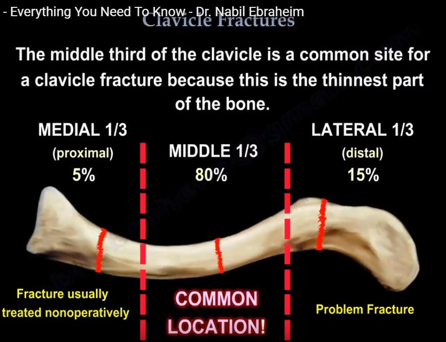
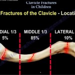
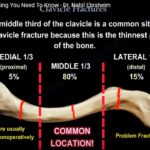
Middle Third (Midshaft): Accounts for approximately 80% of all fractures. It is the thinnest segment and lacks ligamentous reinforcement.
-
Lateral Third: Accounts for roughly 15% of fractures. These are prone to instability due to coracoclavicular ligament disruption.
-
Medial Third: Rare pattern representing roughly 5% of fractures. Most heal predictably with non-operative care.
-
Young Patients with Medial Pain: Always obtain a CT scan. The medial clavicular epiphysis is the last ossification center to fuse in the skeleton (up to age 22 to 25).
| Fracture Location | Incidence (%) | Primary Management Strategy |
| Middle Third | ~80% | Non-operative if non-displaced; ORIF if shortened or comminuted |
| Lateral Third | ~15% | Variable; often requires fixation if ligaments are torn |
| Medial Third | ~5% | Predominantly conservative; CT rule-out for physeal injury |
Conservative Management & Healing Trade-Offs
-
Standard Non-Operative Protocol: Sling immobilization followed by staged physiotherapy.
-
Healing Rate: Conservative treatment achieves an approximate 85% success rate.
-
Callus Formation: Non-operative treatment produces robust secondary bone healing with visible peripheral callus. Rigid plating results in primary healing with minimal to no external callus.
-
Non-Union & Malunion Risks:
-
Non-union develops in roughly 15% of conservatively treated cases.
-
Symptomatic malunion occurs in approximately 10% of cases.
-
Displacement greater than 1.5 cm increases the non-union rate up to 30%.
-
-
Functional Deficits: Symptomatic malunion or non-union may reduce shoulder girdle endurance and peak muscle strength.
-
Range of Motion: Long-term total active shoulder motion is essentially equivalent between operative and non-operative groups.
Clinical Evaluation & Critical Anatomy
-
Inspection: Look for localized deformity, focal ecchymosis, and palpable subcutaneous prominence.
-
Skin Tenting: Marked displacement stretches the overlying skin, creating a risk of open conversion or necrosis.
-
Neurovascular Assessment:
-
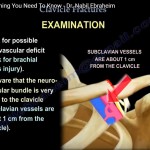
The brachial plexus and subclavian vessels course directly inferior to the middle third of the clavicle.
-
High-yield anatomical relationship: The posterior cord of the brachial plexus is closely related to midshaft fragments.
-
Innervation pearl: The clavicular head of the pectoralis major is innervated specifically by the lateral pectoral nerve.
-
Indications for Surgical Fixation
-
Fracture displacement exceeding 100% (no cortical contact).
-
Shortening greater than 2.0 cm on panoramic bilateral shoulder views.
-
Severe segmental comminution.
-
Z-Type Fractures: Vertical or oblique intermediate fragments resemble the letter Z.
-
The spike can penetrate downward toward the brachial plexus and subclavian vessels.
-
Fixing this pattern acutely avoids hazardous neurovascular dissection during late non-union surgery.
-
| Clinical Parameter | Operative Threshold |
| Cortical Displacement | Greater than 100% translation |
| Bony Shortening | Greater than 2.0 cm |
| Comminution Pattern | Segmental or vertical Z-type fragment |
| Skin Integrity | Threatened soft tissue envelope (tenting) |
Surgical Plating Techniques & Complications
-
Construct Design: Pre-contoured anatomical clavicle plates. Locking screws are reserved for osteopenic bone; non-locking screws are standard in young bone.
-
Plate Position Options:
-
Superior: Stronger biomechanical resistance against inferior bending forces.
-
Anteroinferior: Reduces hardware prominence and lowers the risk of superior screw penetration into the chest.
-
-
Symptomatic Hardware: Hardware irritation is common. Roughly 30% of patients require a second surgery for plate removal after complete healing.
-
Supraclavicular Nerve Injury:
-
Two to three cutaneous branches cross the clavicle in 97% of individuals.
-
More than 50% of operative patients develop numbness in the infraclavicular zone.
-
Numbness can persist past 1 year postoperatively.
-
Safe zones extend 2.0 cm from the sternoclavicular joint and 2.0 cm from the acromioclavicular joint, but these windows are too small for plate placement.
-
-
Number Needed to Treat (NNT): Surgeons operate on roughly seven patients with displaced clavicle fractures to prevent a single non-union.
Management of Clavicular Non-Union
-
Diagnostic Imaging: Plain radiographs can hide non-unions due to cortical overlap. Obtain a CT scan with multiplanar reconstructions when non-union is suspected.
-
Dual Requirements for Healing:
-
Biological Stimulation: Provide local autograft, iliac crest bone graft, or osteoinductive bone graft substitutes.
-
Mechanical Stability: Rigid fixation using a superior pre-contoured plate engaging a minimum of three bicortical screws (six cortices) on each side of the non-union site.
-
Leave a Reply